
Application
Western blot analysis of CEBPB Antibody (N-term) in Hela cell line lysates (35ug/lane). CEBPB (arrow) was detected using the purified Pab.Application
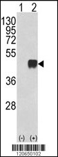
Western blot analysis of CEBPB (arrow) using rabbit polyclonal CEBPB Antibody (N-term) . 293 cell lysates (2 ug/lane) either nontransfected (Lane 1) or transiently transfected with the CEBPB gene (Lane 2) .Application
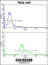
Flow cytometric analysis of hela cells using CEBPB Antibody (N-term)(bottom histogram) compared to a negative control cell (top histogram)FITC-conjugated goat-anti-rabbit secondary antibodies were used for the analysis.Application
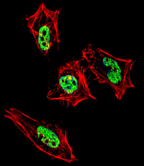
Fluorescent confocal image of Hela cell stained with CEBPB Antibody (N-term)(Cat#AP6815a). Hela cells were fixed with 4% PFA (20 min), permeabilized with Triton X-100 (0.1%, 10 min), then incubated with CEBPB primary antibody (1:25, 1 h at 37℃). For secondary antibody, Alexa Fluor® 488 conjugated donkey anti-rabbit antibody (green) was used (1:400, 50 min at 37℃).Cytoplasmic actin was counterstained with Alexa Fluor® 555 (red) conjugated Phalloidin (7units/ml, 1 h at 37℃). Nuclei were counterstained with DAPI (blue) (10 µg/ml, 10 min). CEBPB immunoreactivity is localized to nucleus significantly.| Product Name | CEBPB Antibody (N-term) |
|---|---|
| Antibody Type | Primary Antibodies |
| Antigen Alias | CEBPB; LAP; TCF5; CCAAT/enhancer-binding protein beta; Liver activator protein; Nuclear factor NF-IL6; Transcription factor 5 |
| Clonality | Polyclonal |
|---|---|
| Isotype | Ig |
| Host Species | Rabbit |
| Tested Applications | WBFCIF |
| WB:1:50~100 FC |
|
| Species Reactivity | Human |
| Concentration | 1mg/ml |
| Gene Synonyms | LAP TCF5 |
|---|---|
| Alternative Names | CEBPB LAP TCF5 CCAAT/enhancer-binding protein beta Liver activator protein Nuclear factor NF-IL6 Transcription factor 5 |
| Molecular Weight(MW) | 36106 Da |
| Function | Important transcriptional activator in the regulation of genes involved in immune and inflammatory responses. Specifically binds to an IL-1 response element in the IL-6 gene. NF-IL6 also binds to regulatory regions of several acute-phase and cytokines genes. It probably plays a role in the regulation of acute-phase reaction, inflammation and hemopoiesis. The consensus recognition site is 5'-T[TG]NNGNAA[TG]-3'. Functions in brown adipose tissue (BAT) differentiation (By similarity). Regulates the transcriptional induction of peroxisome proliferator-activated receptor gamma (PPARG) |
| Tissue Specificity | Expressed at low levels in the lung, kidney and spleen |
| Cellular Localization | Nucleus. |
| Entrez Gene | 1051 |
|---|
Application
Western blot analysis of CEBPB Antibody (N-term) in Hela cell line lysates (35ug/lane). CEBPB (arrow) was detected using the purified Pab.
Application
Western blot analysis of CEBPB (arrow) using rabbit polyclonal CEBPB Antibody (N-term) . 293 cell lysates (2 ug/lane) either nontransfected (Lane 1) or transiently transfected with the CEBPB gene (Lane 2) .
Application
Flow cytometric analysis of hela cells using CEBPB Antibody (N-term)(bottom histogram) compared to a negative control cell (top histogram)FITC-conjugated goat-anti-rabbit secondary antibodies were used for the analysis.
Application
Fluorescent confocal image of Hela cell stained with CEBPB Antibody (N-term)(Cat#AP6815a). Hela cells were fixed with 4% PFA (20 min), permeabilized with Triton X-100 (0.1%, 10 min), then incubated with CEBPB primary antibody (1:25, 1 h at 37℃). For secondary antibody, Alexa Fluor® 488 conjugated donkey anti-rabbit antibody (green) was used (1:400, 50 min at 37℃).Cytoplasmic actin was counterstained with Alexa Fluor® 555 (red) conjugated Phalloidin (7units/ml, 1 h at 37℃). Nuclei were counterstained with DAPI (blue) (10 µg/ml, 10 min). CEBPB immunoreactivity is localized to nucleus significantly.| Application Notes | WB:1:50~100 FC |
|---|
| Form | Liquid |
|---|---|
| Storage Instructions | For short-term storage, store at 4° C. For long-term storage, aliquot and store at -20ºC or below. Avoid multiple freeze-thaw cycles. |
| Storage Buffer | Purified polyclonal antibody supplied in PBS with 0.09% (W/V) sodium azide. This antibody is prepared by Saturated Ammonium Sulfate (SAS) precipitation followed by dialysis against PBS. |


 Data sheet for OM229545