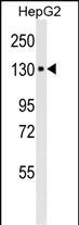
WB
Western blot analysis of anti-ITA6 in HepG2 cell line lysates (35μg/lane). ITA6 (arrow) was detected using the purified Mab.(8μg/ml)IHC
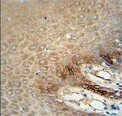
ITA6 Monoclonal Antibody immunohistochemistry analysis in formalin fixed and paraffin embedded human skin carcinoma followed by peroxidase conjugation of the secondary antibody and DAB staining. This data demonstrates the use of the ITA6 Monoclonal Antibody for immunohistochemistry. Clinical relevance has not been evaluated.ICC/IF
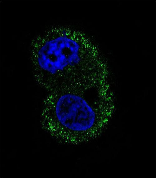
Confocal immunofluorescent analysis of ITA6 Antibody with HepG2 cell followed by Alexa Fluor® 488-conjugated goat anti-mouse lgG (green). DAPI was used to stain the cell nuclear (blue).FC
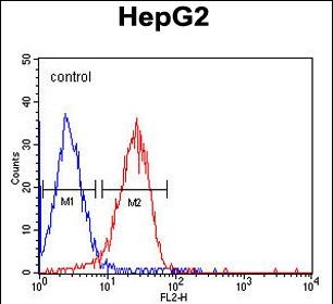
ITA6 Monoclonal Antibody flow cytometric analysis of HepG2 cells (right histogram) compared to a negative control cell (left histogram).PE-conjugated goat-anti-mouse secondary antibodies were used for the analysis.| Product Name | Mouse Monoclonal Antibody to ITA6 |
|---|---|
| Antibody Type | Primary Antibodies |
| Immunogen | ITA6 recombinant protein is used to produce this monoclonal antibody. |
| Clonality | monoclonal |
|---|---|
| Isotype | IgG1 |
| Host Species | Mouse |
| Tested Applications | FCICC/IFIHCWB |
| WB:1:100-1:2000 IHC:1:100-1:500 ICC/IF:1:10-1:50 FC:1:10-1:50 |
|
| Species Reactivity | HumanMouse |
| Concentration | 1mg/ml |
| Purification | Affinity purified |
| Gene Symbol | ITGA6 |
|---|---|
| Gene Synonyms | JEB6 CD49f VLA-6 ITGA6A ITGA6B |
| Gene Full Name | integrin subunit alpha 6 |
| Gene Summary | The gene encodes a member of the integrin alpha chain family of proteins. Integrins are heterodimeric integral membrane proteins composed of an alpha chain and a beta chain that function in cell surface adhesion and signaling. The encoded preproprotein is proteolytically processed to generate light and heavy chains that comprise the alpha 6 subunit. This subunit may associate with a beta 1 or beta 4 subunit to form an integrin that interacts with extracellular matrix proteins including members of the laminin family. The alpha 6 beta 4 integrin may promote tumorigenesis, while the alpha 6 beta 1 integrin may negatively regulate erbB2/HER2 signaling. Alternative splicing results in multiple transcript variants. [provided by RefSeq, Oct 2015] |
| Molecular Weight(MW) | 126kDa |
| Cellular Localization | Cell membrane. |
| Entrez Gene | 3655 |
|---|
WB
Western blot analysis of anti-ITA6 in HepG2 cell line lysates (35μg/lane). ITA6 (arrow) was detected using the purified Mab.(8μg/ml)
IHC
ITA6 Monoclonal Antibody immunohistochemistry analysis in formalin fixed and paraffin embedded human skin carcinoma followed by peroxidase conjugation of the secondary antibody and DAB staining. This data demonstrates the use of the ITA6 Monoclonal Antibody for immunohistochemistry. Clinical relevance has not been evaluated.
ICC/IF
Confocal immunofluorescent analysis of ITA6 Antibody with HepG2 cell followed by Alexa Fluor® 488-conjugated goat anti-mouse lgG (green). DAPI was used to stain the cell nuclear (blue).
FC
ITA6 Monoclonal Antibody flow cytometric analysis of HepG2 cells (right histogram) compared to a negative control cell (left histogram).PE-conjugated goat-anti-mouse secondary antibodies were used for the analysis.| Application Notes | WB:1:100-1:2000 IHC:1:100-1:500 ICC/IF:1:10-1:50 FC:1:10-1:50 |
|---|
| Form | Liquid |
|---|---|
| Storage Instructions | Store at 4°C short term. Aliquot and store at -20°C long term. Avoid freeze/thaw cycles. |
| Storage Buffer | Purified antibody in TBS with 0.05% sodium azide. |


 Data sheet for OM236153