WB
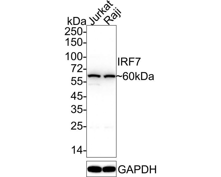
Western blot analysis of IRF7 on different lysates with Rabbit anti-IRF7 antibody at 1/1,000 dilution. Lane 1: Jurkat cell lysate, Lane 2: Raji cell lysate, Lysates/proteins at 15 µg/Lane. Exposure time: 10 seconds; 4-20% SDS-PAGE gel. Proteins were transferred to a PVDF membrane and blocked with 5% NFDM/TBST for 1 hour at room temperature. The primary antibody at 1/1,000 dilution was used in 5% NFDM/TBST at 4℃ overnight. Goat Anti-Rabbit IgG - HRP Secondary Antibody at 1/50,000 dilution was used for 1 hour at room temperature.ICC/IF
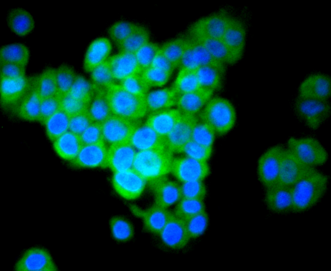
ICC staining IRF7 in PC-12 cells (green). The nuclear counter stain is DAPI (blue). Cells were fixed in paraformaldehyde, permeabilised with 0.25% Triton X100/PBS.ICC/IF
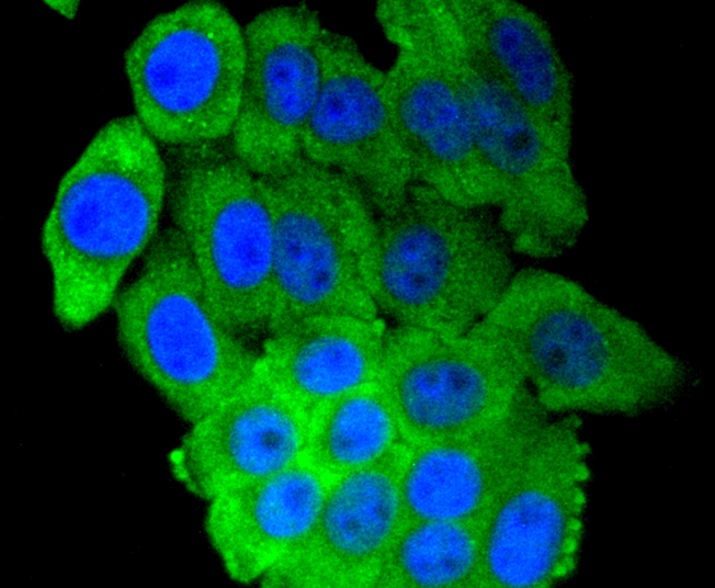
ICC staining IRF7 in HepG2 cells (green). The nuclear counter stain is DAPI (blue). Cells were fixed in paraformaldehyde, permeabilised with 0.25% Triton X100/PBS.ICC/IF
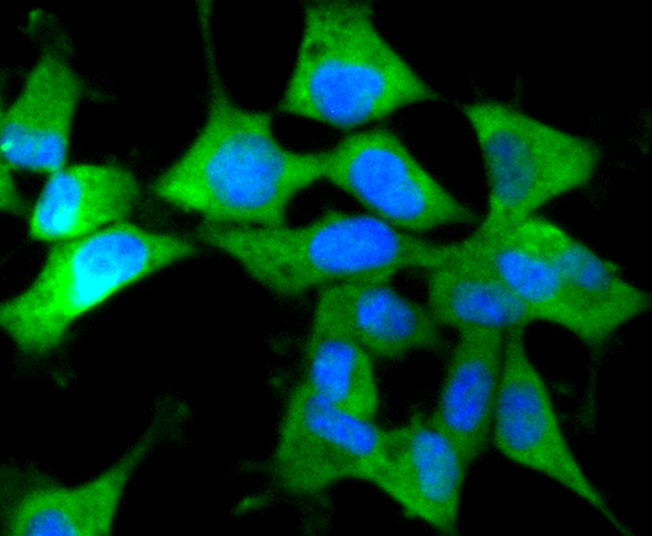
ICC staining IRF7 in 293 cells (green). The nuclear counter stain is DAPI (blue). Cells were fixed in paraformaldehyde, permeabilised with 0.25% Triton X100/PBS.IHC
Immunohistochemical analysis of paraffin-embedded human tonsil tissue using anti-IRF7 antibody. Counter stained with hematoxylin.IHC
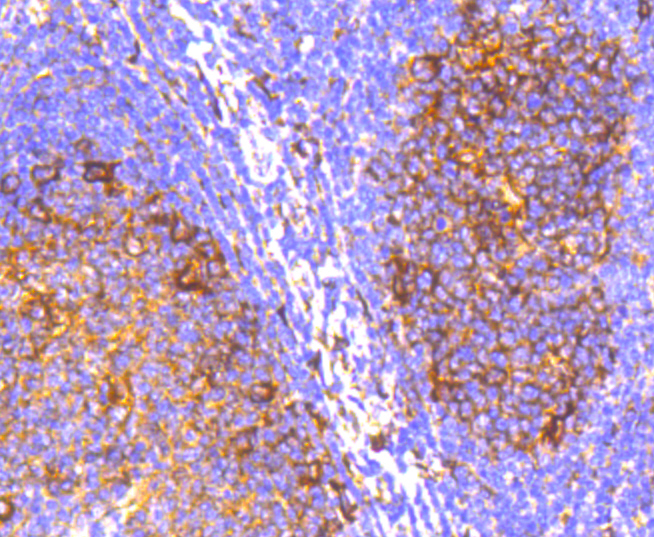
Immunohistochemical analysis of paraffin-embedded human spleen tissue using anti-IRF7 antibody. Counter stained with hematoxylin.IHC
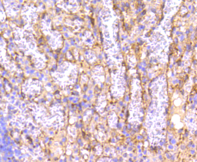
Immunohistochemical analysis of paraffin-embedded human kidney tissue using anti-IRF7 antibody. Counter stained with hematoxylin.IHC
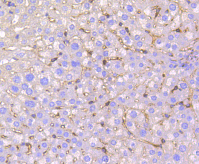
Immunohistochemical analysis of paraffin-embedded mouse liver tissue using anti-IRF7 antibody. Counter stained with hematoxylin.IHC
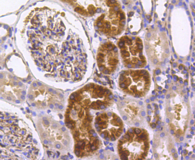
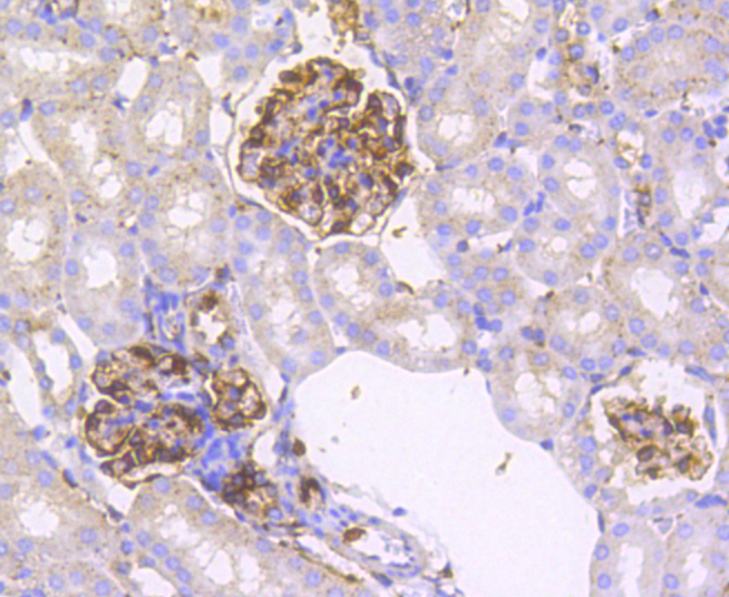
Immunohistochemical analysis of paraffin-embedded mouse kidney tissue using anti-IRF7 antibody. Counter stained with hematoxylin.IHC
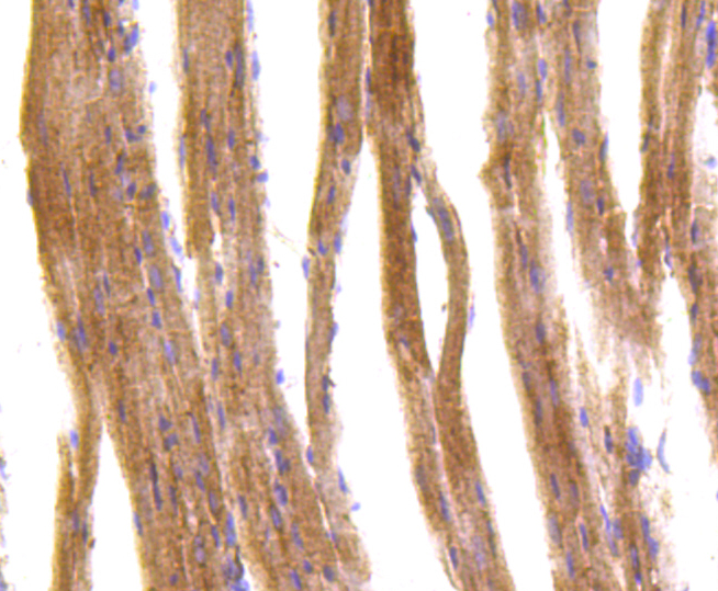
Immunohistochemical analysis of paraffin-embedded mouse heart tissue using anti-IRF7 antibody. Counter stained with hematoxylin.FC
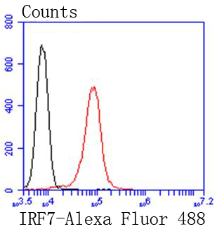
Flow cytometric analysis of Jurkat cells with IRF7 antibody at 1/50 dilution (red) compared with an unlabelled control (cells without incubation with primary antibody; black). Alexa Fluor 488-conjugated goat anti rabbit IgG was used as the secondary antibody| Product Name | IRF7 Recombinant Rabbit Monoclonal Antibody |
|---|---|
| Antibody Type | Primary Antibodies |
| Immunogen | Synthetic peptide within human IRF7 aa 200-240. |
| Clonality | monoclonal |
|---|---|
| Isotype | IgG |
| Host Species | Recombinant rabbit |
| Tested Applications | FCICC/IFIHCIPWB |
| WB:1:2000-1:5000 ICC/IF:1:100-1:500 IHC:1:50-1:200 FC:1:100 |
|
| Species Reactivity | HumanMouseRat |
| Concentration | 1mg/ |
| Purification | Protein A |
| Gene Symbol | IRF7 |
|---|---|
| Gene Synonyms | IMD39 IRF-7 IRF7A IRF7B IRF7C IRF7H IRF-7H |
| Gene Full Name | interferon regulatory factor 7 |
| Gene Summary | This gene encodes interferon regulatory factor 7, a member of the interferon regulatory transcription factor (IRF) family. It has been shown to play a role in the transcriptional activation of virus-inducible cellular genes, including interferon beta chain genes. Inducible expression of IRF7 is largely restricted to lymphoid tissue. The encoded protein plays an important role in the innate immune response against DNA and RNA viruses. [provided by RefSeq, Jul 2021] |
| Molecular Weight(MW) | 54kDa(Observed band size: 60kDa) |
| Cellular Localization | Nucleus, Cytoplasm. |
WB
Western blot analysis of IRF7 on different lysates with Rabbit anti-IRF7 antibody at 1/1,000 dilution. Lane 1: Jurkat cell lysate, Lane 2: Raji cell lysate, Lysates/proteins at 15 µg/Lane. Exposure time: 10 seconds; 4-20% SDS-PAGE gel. Proteins were transferred to a PVDF membrane and blocked with 5% NFDM/TBST for 1 hour at room temperature. The primary antibody at 1/1,000 dilution was used in 5% NFDM/TBST at 4℃ overnight. Goat Anti-Rabbit IgG - HRP Secondary Antibody at 1/50,000 dilution was used for 1 hour at room temperature.
ICC/IF
ICC staining IRF7 in PC-12 cells (green). The nuclear counter stain is DAPI (blue). Cells were fixed in paraformaldehyde, permeabilised with 0.25% Triton X100/PBS.
ICC/IF
ICC staining IRF7 in HepG2 cells (green). The nuclear counter stain is DAPI (blue). Cells were fixed in paraformaldehyde, permeabilised with 0.25% Triton X100/PBS.
ICC/IF
ICC staining IRF7 in 293 cells (green). The nuclear counter stain is DAPI (blue). Cells were fixed in paraformaldehyde, permeabilised with 0.25% Triton X100/PBS.
IHC
Immunohistochemical analysis of paraffin-embedded human tonsil tissue using anti-IRF7 antibody. Counter stained with hematoxylin.
IHC
Immunohistochemical analysis of paraffin-embedded human spleen tissue using anti-IRF7 antibody. Counter stained with hematoxylin.
IHC
Immunohistochemical analysis of paraffin-embedded human kidney tissue using anti-IRF7 antibody. Counter stained with hematoxylin.
IHC
Immunohistochemical analysis of paraffin-embedded mouse liver tissue using anti-IRF7 antibody. Counter stained with hematoxylin.
IHC
Immunohistochemical analysis of paraffin-embedded mouse kidney tissue using anti-IRF7 antibody. Counter stained with hematoxylin.
IHC
Immunohistochemical analysis of paraffin-embedded mouse heart tissue using anti-IRF7 antibody. Counter stained with hematoxylin.
FC
Flow cytometric analysis of Jurkat cells with IRF7 antibody at 1/50 dilution (red) compared with an unlabelled control (cells without incubation with primary antibody; black). Alexa Fluor 488-conjugated goat anti rabbit IgG was used as the secondary antibody| Positive Control | Jurkat, Raji, PC-12, HepG2, mouse liver tissue, human tonsil tissue, mouse kidney tissue, human spleen tissue, mouse heart tissue, human kidney tissue. |
|---|---|
| Application Notes | WB:1:2000-1:5000 ICC/IF:1:100-1:500 IHC:1:50-1:200 FC:1:100 |
| Form | Liquid |
|---|---|
| Storage Instructions | Store at +4℃ after thawing. Aliquot store at -20℃ or -80℃. Avoid repeated freeze / thaw cycles. |
| Storage Buffer | 1*TBS (pH7.4), 1%BSA, 40%Glycerol. Preservative: 0.05% Sodium Azide. |


 Data sheet for OM637418