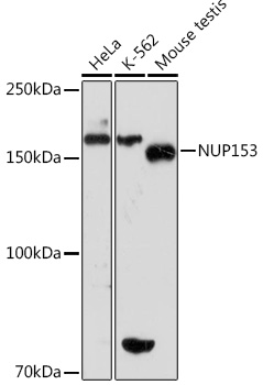


WB
Western blot analysis of various lysates using NUP153 Rabbit mAb at 1:1000 dilution. Secondary antibody: HRP Goat Anti-Rabbit IgG (H+L)at 1:10000 dilution. Lysates/proteins: 25μg per lane. Blocking buffer: 3% nonfat dry milk in TBST. Detection: ECL Basic Kit. Exposure time: 30s.IHC
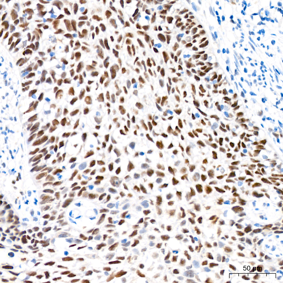
Immunohistochemistry analysis of NUP153 in paraffin-embedded human cervix cancer tissue using NUP153 Rabbit mAb at a dilution of 1:200 (40x lens).High pressure antigen retrieval was performed with 0.01 M citrate buffer (pH 6.0) prior to IHC staining.IHC
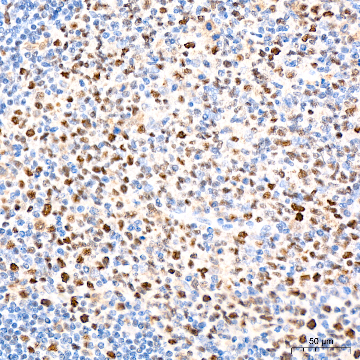
Immunohistochemistry analysis of NUP153 in paraffin-embedded human tonsil tissue using NUP153 Rabbit mAb at a dilution of 1:200 (40x lens).High pressure antigen retrieval was performed with 0.01 M citrate buffer (pH 6.0) prior to IHC staining.IHC
Immunohistochemistry analysis of NUP153 in paraffin-embedded mouse testis tissue using NUP153 Rabbit mAb at a dilution of 1:200 (40x lens).High pressure antigen retrieval was performed with 0.01 M citrate buffer (pH 6.0) prior to IHC staining.ICC/IF
Immunofluorescence analysis of U-2 OS cells using NUP153 Rabbit mAb at dilution of 1:100 (40x lens). Secondary antibody: Cy3 Goat Anti-Rabbit IgG (H+L) at 1:500 dilution. Blue: DAPI for nuclear staining.| Product Name | NUP153 Rabbit mAb |
|---|---|
| Antibody Type | Primary Antibodies |
| Immunogen | A synthetic peptide corresponding to a sequence within amino acids 1-100 of human NUP153 (P49790). |
| Clonality | monoclonal |
|---|---|
| Isotype | IgG |
| Host Species | Rabbit |
| Tested Applications | ICC/IFIHCWB |
| WB:1:500-1:1000 IHC:1:50-1:200 ICC/IF:1:50-1:200 |
|
| Species Reactivity | HumanMouseRat |
| Concentration | 1mg/ml |
| Purification | Affinity purified |
| Gene Symbol | NUP153 |
|---|---|
| Gene Synonyms | N153 HNUP153 |
| Gene Full Name | nucleoporin 153 |
| Gene Summary | Nuclear pore complexes regulate the transport of macromolecules between the nucleus and cytoplasm. They are composed of at least 100 different polypeptide subunits, many of which belong to the nucleoporin family. Nucleoporins are glycoproteins found in nuclear pores and contain characteristic pentapeptide XFXFG repeats as well as O-linked N-acetylglucosamine residues oriented towards the cytoplasm. The protein encoded by this gene has three distinct domains: a N-terminal region containing a pore targeting and an RNA-binding domain domain, a central region containing multiple zinc finger motifs, and a C-terminal region containing multiple XFXFG repeats. Alternative splicing results in multiple transcript variants of this gene. [provided by RefSeq, May 2013] |
| Molecular Weight(MW) | 154kDa |
| Cellular Localization | Nucleus, Nucleus membrane, nuclear pore complex. |
WB
Western blot analysis of various lysates using NUP153 Rabbit mAb at 1:1000 dilution. Secondary antibody: HRP Goat Anti-Rabbit IgG (H+L)at 1:10000 dilution. Lysates/proteins: 25μg per lane. Blocking buffer: 3% nonfat dry milk in TBST. Detection: ECL Basic Kit. Exposure time: 30s.
IHC
Immunohistochemistry analysis of NUP153 in paraffin-embedded human cervix cancer tissue using NUP153 Rabbit mAb at a dilution of 1:200 (40x lens).High pressure antigen retrieval was performed with 0.01 M citrate buffer (pH 6.0) prior to IHC staining.
IHC
Immunohistochemistry analysis of NUP153 in paraffin-embedded human tonsil tissue using NUP153 Rabbit mAb at a dilution of 1:200 (40x lens).High pressure antigen retrieval was performed with 0.01 M citrate buffer (pH 6.0) prior to IHC staining.
IHC
Immunohistochemistry analysis of NUP153 in paraffin-embedded mouse testis tissue using NUP153 Rabbit mAb at a dilution of 1:200 (40x lens).High pressure antigen retrieval was performed with 0.01 M citrate buffer (pH 6.0) prior to IHC staining.
ICC/IF
Immunofluorescence analysis of U-2 OS cells using NUP153 Rabbit mAb at dilution of 1:100 (40x lens). Secondary antibody: Cy3 Goat Anti-Rabbit IgG (H+L) at 1:500 dilution. Blue: DAPI for nuclear staining.| Application Notes | WB:1:500-1:1000 IHC:1:50-1:200 ICC/IF:1:50-1:200 |
|---|
| Form | Liquid |
|---|---|
| Storage Instructions | Store at -20℃. Avoid freeze / thaw cycles. |
| Storage Buffer | Buffer: PBS with 0.02% sodium azide, 0.05% BSA, 50% glycerol, pH7.3. |


 Data sheet for OM644272