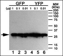
Western blot analysis of anti-GFP Mab (Cat. #AM1009a) using purified GFP, YFP and BFP proteins expressed in bacteria: Both GFP (Lanes 1-3) and YFP (Lanes 4-6) but not BFP (data not shown) were detected using the purified Mab.

Western blot analysis of anti-GFP Tag Antibody (Cat. #AM1009a) in GFP recombinant protein. GFP recombinant protein (arrow) was detected using the purified Mab.

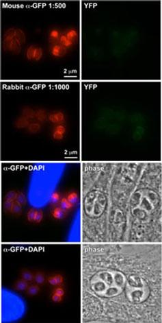
Immuno-fluorescence for GFP Tag Antibody (Cat # AM1009A). Antibody target is a YFP tagged protein (the inner membrane complex) of Toxoplasma gondii, which localizes around the exterior of the parasite. Brighter parasites represent actively dividing cells and darker ones are not actively dividing.Secondary goat-anti-mouse and goat-anti-rabbit were conjugated with Alexa546 (Molecular Probes)at 1:200 dilution. Phase contrast enhancement applied. Fluorescent lamp exposure times as follows: RFP Channel, 0.075 sec; YFP Channel, 1.000 sec; DAPI Channel, 0.020 sec; Phase Channel: 0.300 sec. Microscope: Axiovert 200M inverted microscope (Zeiss) equipped with a PRIOR stage and a NEOFLUAR 100x/1.3 objective lens. Images were collected with a Hamamatsu C4742-95 CCD camera. Chroma Filtersets: TRITC, HQ545/30x – Q570LP – HQ620/60m; DAPI, D350/50x – E420LP – 400DCLP; YFP, Q151LP – HQ500/20x – HQ535/30m. Data courtesy of Seth Robertson, Boston College.

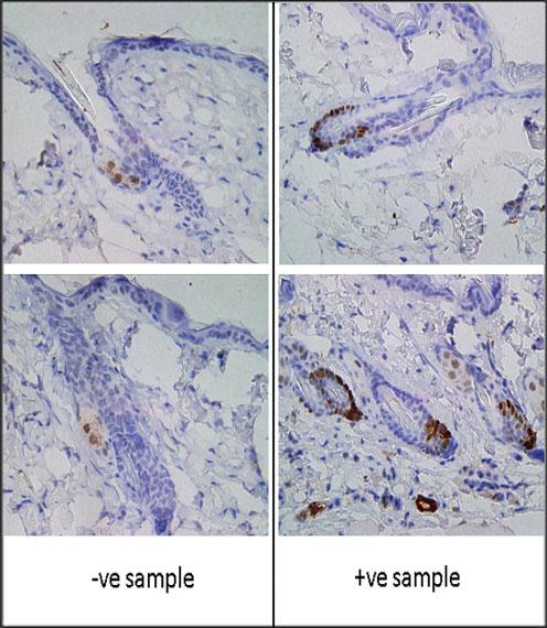
Formalin-fixed and paraffin-embedded mouse skin tissue expression GFP reacted with GFP Tag Antibody (Cat.#AM1009a), which was peroxidase-conjugated to the secondary antibody, followed by DAB staining. -ve shows negative staining of the antibody in a wild type mouse, whereas +ve shows positive staining in a mouse expressing GFP under the LGR5 promoter. The staining shows localization of GFP at the bottom of the hair follicle. The antigen was retrieved in citrate pH 6 in water bath for 25 mins.
Product Profile
| Product Name | GFP Tag Antibody |
|---|---|
| Antibody Type | Tags Antibodies |
Key Feature
| Clonality | Monoclonal |
|---|---|
| Isotype | IgG1 |
| Clone Number | 168AT1211 |
| Host Species | Mouse |
| Tested Applications | |
WB~~1:2,000 IF~~1:50~100 IHC~~1:50~100: |
|
| Species Reactivity | |
| Concentration | 1 mg/ml |
| Purification |
Target Information
| Molecular Weight(MW) | 48 kDa |
|---|---|
| Tissue Specificity | Purified His-tagged GFP protein was used to produced this monoclonal antibody. |
Application
-

Application
Western blot analysis of anti-GFP Mab (Cat. #AM1009a) using purified GFP, YFP and BFP proteins expressed in bacteria: Both GFP (Lanes 1-3) and YFP (Lanes 4-6) but not BFP (data not shown) were detected using the purified Mab.
-

Application
Western blot analysis of anti-GFP Tag Antibody (Cat. #AM1009a) in GFP recombinant protein. GFP recombinant protein (arrow) was detected using the purified Mab.
-

Application
Immuno-fluorescence for GFP Tag Antibody (Cat # AM1009A). Antibody target is a YFP tagged protein (the inner membrane complex) of Toxoplasma gondii, which localizes around the exterior of the parasite. Brighter parasites represent actively dividing cells and darker ones are not actively dividing.Secondary goat-anti-mouse and goat-anti-rabbit were conjugated with Alexa546 (Molecular Probes)at 1:200 dilution. Phase contrast enhancement applied. Fluorescent lamp exposure times as follows: RFP Channel, 0.075 sec; YFP Channel, 1.000 sec; DAPI Channel, 0.020 sec; Phase Channel: 0.300 sec. Microscope: Axiovert 200M inverted microscope (Zeiss) equipped with a PRIOR stage and a NEOFLUAR 100x/1.3 objective lens. Images were collected with a Hamamatsu C4742-95 CCD camera. Chroma Filtersets: TRITC, HQ545/30x – Q570LP – HQ620/60m; DAPI, D350/50x – E420LP – 400DCLP; YFP, Q151LP – HQ500/20x – HQ535/30m. Data courtesy of Seth Robertson, Boston College.
-

Application
Formalin-fixed and paraffin-embedded mouse skin tissue expression GFP reacted with GFP Tag Antibody (Cat.#AM1009a), which was peroxidase-conjugated to the secondary antibody, followed by DAB staining. -ve shows negative staining of the antibody in a wild type mouse, whereas +ve shows positive staining in a mouse expressing GFP under the LGR5 promoter. The staining shows localization of GFP at the bottom of the hair follicle. The antigen was retrieved in citrate pH 6 in water bath for 25 mins.
| Application Notes | WB~~1:2,000 IF~~1:50~100 IHC~~1:50~100: |
|---|
Additional Information
| Form | Liquid |
|---|---|
| Storage Instructions | For short-term storage, store at 4° C. For long-term storage, aliquot and store at -20ºC or below. Avoid multiple freeze-thaw cycles. |
| Storage Buffer | Purified monoclonal antibody supplied in PBS with 0.09% (W/V) sodium azide. This antibody is purified by protein G and Ni-NTA nickel affinity chromatography, followed by dialysis against PBS. |
- Related products
- Antigen Repair 20X pH6.0 OM642703
- 647 labeled Tyramide OM642699
- 594 labeled Tyramide OM642698
- 555 labeled Tyramide OM642697
- 525 labeled Tyramide OM642696
-
- ASSAY KITS
-
- SERUM
2013 © Omnimabs , All Rights Reserved.

