Application
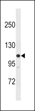
MLCK Antibody (M1) western blot analysis in SK-BR-3 cell line lysates (35ug/lane).This demonstrates the MLCK antibody detected the MLCK protein (arrow).| Product Name | MLCK Antibody (N-term) |
|---|---|
| Antibody Type | Primary Antibodies |
| Antigen Alias | Myosin light chain kinase |
| Clonality | Polyclonal |
|---|---|
| Isotype | Ig |
| Host Species | Rabbit |
| Tested Applications | WB |
| WB:1:100~500 |
|
| Species Reactivity | Human |
| Concentration | 1mg/ml |
| Gene Synonyms | MLCK MLCK1 MYLK1 |
|---|---|
| Alternative Names | Myosin light chain kinase |
| Molecular Weight(MW) | 210715 Da |
| Function | Calcium/calmodulin-dependent myosin light chain kinase implicated in smooth muscle contraction via phosphorylation of myosin light chains (MLC). Also regulates actin-myosin interaction through a non-kinase activity. Phosphorylates PTK2B/PYK2 and myosin light-chains. Involved in the inflammatory response (e.g apoptosis, vascular permeability, leukocyte diapedesis), cell motility and morphology, airway hyperreactivity and other activities relevant to asthma. Required for tonic airway smooth muscle contraction that is necessary for physiological and asthmatic airway resistance. Necessary for gastrointestinal motility. Implicated in the regulation of endothelial as well as vascular permeability, probably via the regulation of cytoskeletal rearrangements. In the nervous system it has been shown to control the growth initiation of astrocytic processes in culture and to participate in transmitter release at synapses formed between cultured sympathetic ganglion cells. Critical participant in signaling sequences that result in fibroblast apoptosis. Plays a role in the regulation of epithelial cell survival. Required for epithelial wound healing, especially during actomyosin ring contraction during purse-string wound closure. Mediates RhoA- dependent membrane blebbing. Triggers TRPC5 channel activity in a calcium-dependent signaling, by inducing its subcellular localization at the plasma membrane. Promotes cell migration (including tumor cells) and tumor metastasis. PTK2B/PYK2 activation by phosphorylation mediates ITGB2 activation and is thus essential to trigger neutrophil transmigration during acute lung injury (ALI). May regulate optic nerve head astrocyte migration. Probably involved in mitotic cytoskeletal regulation Regulates tight junction probably by modulating ZO-1 exchange in the perijunctional actomyosin ring. Mediates burn-induced microvascular barrier injury; triggers endothelial contraction in the development of microvascular hyperpermeability by phosphorylating MLC. Essential for intestinal barrier dysfunction Mediates Giardia spp.-mediated reduced epithelial barrier function during giardiasis intestinal infection via reorganization of cytoskeletal F-actin and tight junctional ZO-1. Necessary for hypotonicity-induced Ca(2+) entry and subsequent activation of volume-sensitive organic osmolyte/anion channels (VSOAC) in cervical cancer cells. Responsible for high proliferative ability of breast cancer cells through anti-apoptosis |
| Tissue Specificity | Smooth muscle and non-muscle isozymes are expressed in a wide variety of adult and fetal tissues and in cultured endothelium with qualitative expression appearing to be neither tissue- nor development-specific. Non-muscle isoform 2 is the dominant splice variant expressed in various tissues. Telokin has been found in a wide variety of adult and fetal tissues Accumulates in well differentiated enterocytes of the intestinal epithelium in response to tumor necrosis factor (TNF) |
| Cellular Localization | Cytoplasm. Cell projection, lamellipodium. Cleavage furrow. Cytoplasm, cytoskeleton. Note=Localized to stress fibers during interphase and to the cleavage furrow during mitosis |
| Entrez Gene | 4638 |
|---|
Application
MLCK Antibody (M1) western blot analysis in SK-BR-3 cell line lysates (35ug/lane).This demonstrates the MLCK antibody detected the MLCK protein (arrow).| Application Notes | WB:1:100~500 |
|---|
| Form | Liquid |
|---|---|
| Storage Instructions | For short-term storage, store at 4° C. For long-term storage, aliquot and store at -20ºC or below. Avoid multiple freeze-thaw cycles. |
| Storage Buffer | Purified polyclonal antibody supplied in PBS with 0.09% (W/V) sodium azide. This antibody is purified through a protein G column, eluted with high and low pH buffers and neutralized immediately, followed by dialysis against PBS. |


 Data sheet for OM213787