
Application
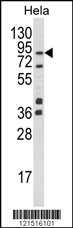
Western blot analysis of UHRF1 Antibody (Center) in Hela cell line lysates (35ug/lane). UHRF1 (arrow) was detected using the purified Pab.Application
UHRF1 Antibody (Center) flow cytometry analysis of Hela cells (bottom histogram) compared to a negative control cell (top histogram). FITC-conjugated goat-anti-rabbit secondary antibodies were used for the analysis.Application
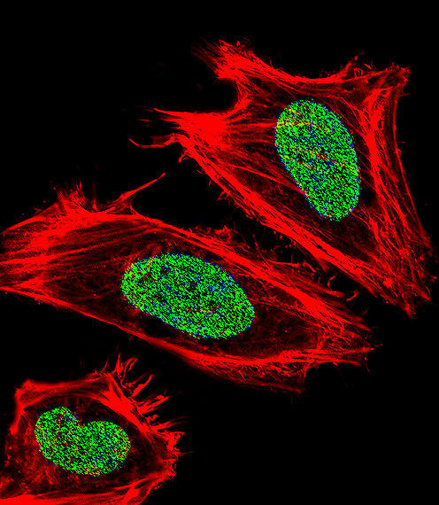
Fluorescent confocal image of Hela cell stained with UHRF1 Antibody (Center)(Cat#AP8846c).Hela cells were fixed with 4% PFA (20 min), permeabilized with Triton X-100 (0.1%, 10 min), then incubated with UHRF1 primary antibody (1:25, 1 h at 37℃). For secondary antibody, Alexa Fluor® 488 conjugated donkey anti-rabbit antibody (green) was used (1:400, 50 min at 37℃).Cytoplasmic actin was counterstained with Alexa Fluor® 555 (red) conjugated Phalloidin (7units/ml, 1 h at 37℃). Nuclei were counterstained with DAPI (blue) (10 µg/ml, 10 min). UHRF1 immunoreactivity is localized to Nucleus significantly.| Product Name | UHRF1 Antibody (Center) |
|---|---|
| Antibody Type | Primary Antibodies |
| Antigen Alias | UHRF1; ICBP90; NP95; RNF106; E3 ubiquitin-protein ligase UHRF1; Inverted CCAAT box-binding protein of 90 kDa; Nuclear protein 95; Nuclear zinc finger protein Np95; RING finger protein 106; Transcription factor ICBP90; Ubiquitin-like PHD and RING finger dom |
| Clonality | Polyclonal |
|---|---|
| Isotype | Ig |
| Host Species | Rabbit |
| Tested Applications | WBFCIF |
| WB:1:100-500 FC |
|
| Species Reactivity | Human |
| Concentration | 1mg/ml |
| Gene Synonyms | ICBP90 NP95 RNF106 |
|---|---|
| Alternative Names | UHRF1 ICBP90 NP95 RNF106 E3 ubiquitin-protein ligase UHRF1 Inverted CCAAT box-binding protein of 90 kDa Nuclear protein 95 Nuclear zinc finger protein Np95 RING finger protein 106 Transcription factor ICBP90 Ubiquitin-like PHD and RING finger dom |
| Molecular Weight(MW) | 89814 Da |
| Function | Multidomain protein that acts as a key epigenetic regulator by bridging DNA methylation and chromatin modification Specifically recognizes and binds hemimethylated DNA at replication forks via its YDG domain and recruits DNMT1 methyltransferase to ensure faithful propagation of the DNA methylation patterns through DNA replication. In addition to its role in maintenance of DNA methylation, also plays a key role in chromatin modification: through its tudor-like regions and PHD- type zinc fingers, specifically recognizes and binds histone H3 trimethylated at 'Lys-9' (H3K9me3) and unmethylated at 'Arg-2' (H3R2me0), respectively, and recruits chromatin proteins. Enriched in pericentric heterochromatin where it recruits different chromatin modifiers required for this chromatin replication. Also localizes to euchromatic regions where it negatively regulates transcription possibly by impacting DNA methylation and histone modifications. Has E3 ubiquitin-protein ligase activity by mediating the ubiquitination of target proteins such as histone H3 and PML. It is still unclear how E3 ubiquitin-protein ligase activity is related to its role in chromatin in vivo. May be involved in DNA repair |
| Tissue Specificity | Expressed in thymus, bone marrow, testis, lung and heart. Overexpressed in breast cancer |
| Cellular Localization | Nucleus. Note=Localizes to replication foci. Enriched in pericentric heterochromatin. Also localizes to euchromatic regions |
| Entrez Gene | 29128 |
|---|
Application
Western blot analysis of UHRF1 Antibody (Center) in Hela cell line lysates (35ug/lane). UHRF1 (arrow) was detected using the purified Pab.
Application
UHRF1 Antibody (Center) flow cytometry analysis of Hela cells (bottom histogram) compared to a negative control cell (top histogram). FITC-conjugated goat-anti-rabbit secondary antibodies were used for the analysis.
Application
Fluorescent confocal image of Hela cell stained with UHRF1 Antibody (Center)(Cat#AP8846c).Hela cells were fixed with 4% PFA (20 min), permeabilized with Triton X-100 (0.1%, 10 min), then incubated with UHRF1 primary antibody (1:25, 1 h at 37℃). For secondary antibody, Alexa Fluor® 488 conjugated donkey anti-rabbit antibody (green) was used (1:400, 50 min at 37℃).Cytoplasmic actin was counterstained with Alexa Fluor® 555 (red) conjugated Phalloidin (7units/ml, 1 h at 37℃). Nuclei were counterstained with DAPI (blue) (10 µg/ml, 10 min). UHRF1 immunoreactivity is localized to Nucleus significantly.| Application Notes | WB:1:100-500 FC |
|---|
| Form | Liquid |
|---|---|
| Storage Instructions | For short-term storage, store at 4° C. For long-term storage, aliquot and store at -20ºC or below. Avoid multiple freeze-thaw cycles. |
| Storage Buffer | Purified polyclonal antibody supplied in PBS with 0.09% (W/V) sodium azide. This antibody is purified through a protein A column, followed by peptide affinity purification. |


 Data sheet for OM224215