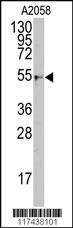
Application
Western blot analysis of anti-KPNA2 Antibody (Center) in A2058 cell line lysates (35ug/lane). KPNA2(arrow) was detected using the purified Pab.Application
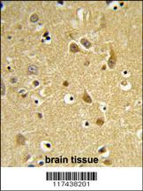
Formalin-fixed and paraffin-embedded human brain tissue reacted with KPNA2 Antibody (Center), which was peroxidase-conjugated to the secondary antibody, followed by DAB staining. This data demonstrates the use of this antibody for immunohistochemistry; clinical relevance has not been evaluated.Application
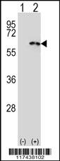
Western blot analysis of KPNA2 (arrow) using rabbit polyclonal KPNA2 Antibody (Center) . 293 cell lysates (2 ug/lane) either nontransfected (Lane 1) or transiently transfected (Lane 2) with the KPNA2 gene.Application
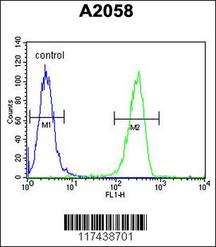
KPNA2 Antibody (Center) flow cytometric analysis of A2058 cells (right histogram) compared to a negative control cell (left histogram).FITC-conjugated goat-anti-rabbit secondary antibodies were used for the analysis.| Product Name | KPNA2 Antibody (Center) |
|---|---|
| Antibody Type | Primary Antibodies |
| Antigen Alias | KPNA2; RCH1; SRP1; Importin subunit alpha-2; Karyopherin subunit alpha-2; RAG cohort protein 1; SRP1-alpha |
| Clonality | Polyclonal |
|---|---|
| Isotype | Ig |
| Host Species | Rabbit |
| Tested Applications | WBIHCFC |
| WB:1:50~100 IHC |
|
| Species Reactivity | Human |
| Concentration | 1mg/ml |
| Gene Synonyms | RCH1 SRP1 |
|---|---|
| Alternative Names | KPNA2 RCH1 SRP1 Importin subunit alpha-2 Karyopherin subunit alpha-2 RAG cohort protein 1 SRP1-alpha |
| Molecular Weight(MW) | 57862 Da |
| Function | Functions in nuclear protein import as an adapter protein for nuclear receptor KPNB1. Binds specifically and directly to substrates containing either a simple or bipartite NLS motif. Docking of the importin/substrate complex to the nuclear pore complex (NPC) is mediated by KPNB1 through binding to nucleoporin FxFG repeats and the complex is subsequently translocated through the pore by an energy requiring, Ran- dependent mechanism. At the nucleoplasmic side of the NPC, Ran binds to importin-beta and the three components separate and importin-alpha and -beta are re-exported from the nucleus to the cytoplasm where GTP hydrolysis releases Ran from importin. The directionality of nuclear import is thought to be conferred by an asymmetric distribution of the GTP- and GDP-bound forms of Ran between the cytoplasm and nucleus |
| Tissue Specificity | Expressed ubiquitously. |
| Cellular Localization | Cytoplasm. Nucleus. |
| Entrez Gene | 3838 |
|---|
Application
Western blot analysis of anti-KPNA2 Antibody (Center) in A2058 cell line lysates (35ug/lane). KPNA2(arrow) was detected using the purified Pab.
Application
Formalin-fixed and paraffin-embedded human brain tissue reacted with KPNA2 Antibody (Center), which was peroxidase-conjugated to the secondary antibody, followed by DAB staining. This data demonstrates the use of this antibody for immunohistochemistry; clinical relevance has not been evaluated.
Application
Western blot analysis of KPNA2 (arrow) using rabbit polyclonal KPNA2 Antibody (Center) . 293 cell lysates (2 ug/lane) either nontransfected (Lane 1) or transiently transfected (Lane 2) with the KPNA2 gene.
Application
KPNA2 Antibody (Center) flow cytometric analysis of A2058 cells (right histogram) compared to a negative control cell (left histogram).FITC-conjugated goat-anti-rabbit secondary antibodies were used for the analysis.| Application Notes | WB:1:50~100 IHC |
|---|
| Form | Liquid |
|---|---|
| Storage Instructions | For short-term storage, store at 4° C. For long-term storage, aliquot and store at -20ºC or below. Avoid multiple freeze-thaw cycles. |
| Storage Buffer | Purified polyclonal antibody supplied in PBS with 0.09% (W/V) sodium azide. This antibody is prepared by Saturated Ammonium Sulfate (SAS) precipitation followed by dialysis against PBS. |


 Data sheet for OM236779