Application
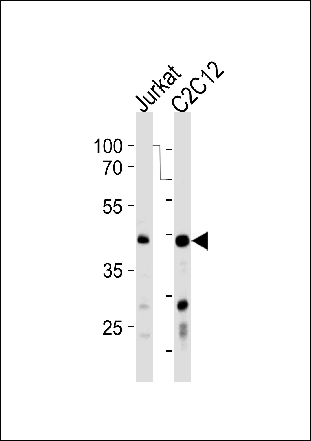
Western blot analysis in Jurkat,mouse C2C12 cell line lysates (35ug/lane).This demonstrates the antibody detected the protein (arrow).Application
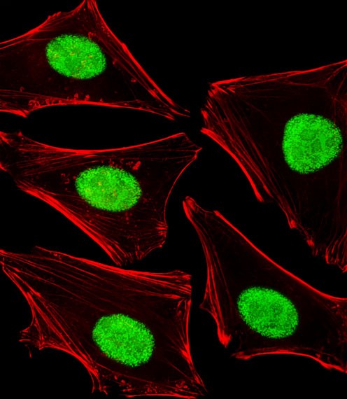
Fluorescent image of Hela cells stained with POLDIP3 Antibody (N-term). Antibody was diluted at 1:25 dilution. An Alexa Fluor 488-conjugated goat anti-rabbit lgG at 1:400 dilution was used as the secondary antibody (green). Cytoplasmic actin was counterstained with Alexa Fluor 555 conjugated with Phalloidin (red).Application
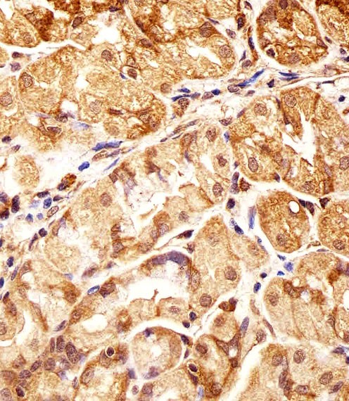
Immunohistochemical analysis of paraffin-embedded H. stomach section using POLDIP3 Antibody (N-term). Antibody was diluted at 1:25 dilution. A undiluted biotinylated goat polyvalent antibody was used as the secondary, followed by DAB staining.| Product Name | POLDIP3 Antibody |
|---|---|
| Antibody Type | Primary Antibodies |
| Antigen Alias | Polymerase delta-interacting protein 3, 46 kDa DNA polymerase delta interaction protein, p46, S6K1 Aly/REF-like target, SKAR, POLDIP3, KIAA1649, PDIP46 |
| Product description | This gene encodes a protein that interacts with the DNA polymerase delta p50 subunit. This protein is a specific target of S6 kinase 1 and regulates cell growth. Two transcript variants that encode different protein isoforms have been identified. [provided by RefSeq].1) Ma, X.M., et al. Cell 133(2):303-313(2008) |
| Immunogen | This POLDIP3 antibody is generated from rabbits immunized with a KLH conjugated synthetic peptide between 93-120 amino acids from the N-terminal region of human POLDIP3. |
| Clonality | Polyclonal |
|---|---|
| Isotype | Ig |
| Host Species | Rabbit |
| Tested Applications | IFIHC-PWB |
| For IHC-P starting dilution is: 1:25 For IF starting dilution is: 1:25 For WB starting dilution is: 1:1000: |
|
| Species Reactivity | HumanMouse |
| Concentration | 1mg/ml |
| Purification | Affinity purified |
| Gene Symbol | POLDIP3 |
|---|---|
| Alternative Names | Polymerase delta-interacting protein 3 46 kDa DNA polymerase delta interaction protein p46 S6K1 Aly/REF-like target SKAR POLDIP3 KIAA1649 PDIP46 |
| Molecular Weight(MW) | 46 kDa |
Application
Western blot analysis in Jurkat,mouse C2C12 cell line lysates (35ug/lane).This demonstrates the antibody detected the protein (arrow).
Application
Fluorescent image of Hela cells stained with POLDIP3 Antibody (N-term). Antibody was diluted at 1:25 dilution. An Alexa Fluor 488-conjugated goat anti-rabbit lgG at 1:400 dilution was used as the secondary antibody (green). Cytoplasmic actin was counterstained with Alexa Fluor 555 conjugated with Phalloidin (red).
Application
Immunohistochemical analysis of paraffin-embedded H. stomach section using POLDIP3 Antibody (N-term). Antibody was diluted at 1:25 dilution. A undiluted biotinylated goat polyvalent antibody was used as the secondary, followed by DAB staining.| Application Notes | For IHC-P starting dilution is: 1:25 For IF starting dilution is: 1:25 For WB starting dilution is: 1:1000: |
|---|
| Form | Liquid |
|---|---|
| Storage Instructions | Store at 4˚C for three months and -20˚C, stable for up to one year. As with all antibodies care should be taken to avoid repeated freeze thaw cycles. Antibodies should not be exposed to prolonged high temperatures. |
| Storage Buffer | Supplied in PBS with 0.09% (W/V) sodium azide. |


 Data sheet for OM288438