Application
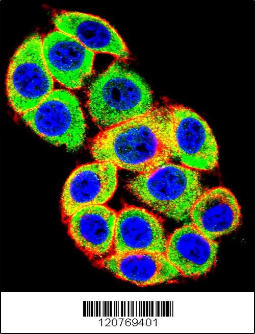
Confocal immunofluorescent analysis of PRKCA Antibody with Hela cell followed by Alexa Fluor 488-conjugated goat anti-rabbit lgG (green). Actin filaments have been labeled with Alexa Fluor 555 phalloidin (red).DAPI was used to stain the cell nuclear (blue).Application
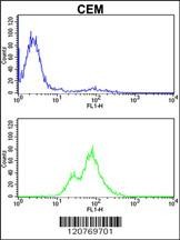
Flow cytometric analysis of CEM cells (bottom histogram) compared to a negative control cell (top histogram).FITC-conjugated goat-anti-rabbit secondary antibodies were used for the analysis.Application
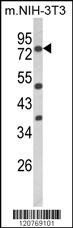
Western blot analysis of PRKCA Antibody in mouse NIH-3T3 cell line lysates (35ug/lane)Application
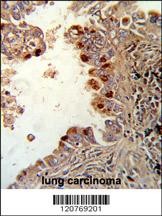
Formalin-fixed and paraffin-embedded human lung carcinoma reacted with PRKCA Antibody (N-term), which was peroxidase-conjugated to the secondary antibody, followed by DAB staining.| Product Name | PRKCA Antibody |
|---|---|
| Antibody Type | Primary Antibodies |
| Product description | PRKCA is a calcium-activated, phospholipid-dependent, serine-and threonine-specific enzyme. It may play a role in cell motility by phosphorylating CSPG4. PKC is activated by diacylglycerol which in turn phosphorylates a range of cellular proteins. PKC also serves as the receptor for phorbol esters, a class of tumor promoters.1) Gysin,S. and Imber,R. et.al., Eur. J. Biochem. 240 (3), 747-750 (1996) Rider,M.H., et.al., Biochem. J. 285 (PT 2), 405-411 (1992) |
| Immunogen | This PRKCA antibody is generated from rabbits immunized with a KLH conjugated synthetic peptide between 75-103 amino acids from the N-terminal region of human PRKCA. |
| Clonality | Polyclonal |
|---|---|
| Isotype | Ig |
| Host Species | Rabbit |
| Tested Applications | FACSIFIHC-PWB |
| For WB starting dilution is: 1:1000 For IHC-P starting dilution is: 1:50~100 For FACS starting dilution is: 1:10~50 For IF starting dilution is: 1:10~50 | |
| Species Reactivity | HumanMouse |
| Concentration | 1mg/ml |
| Purification | Unpurified |
| Gene Symbol | PRKCA |
|---|---|
| Alternative Names | Protein kinase C alpha type PKC-A PKC-alpha PRKCA PKCA PRKACA |
| Molecular Weight(MW) | 77 kDa |
| Sequence Similarities | Predicted species reactivity based on immunogen sequence: Bovine |
Application
Confocal immunofluorescent analysis of PRKCA Antibody with Hela cell followed by Alexa Fluor 488-conjugated goat anti-rabbit lgG (green). Actin filaments have been labeled with Alexa Fluor 555 phalloidin (red).DAPI was used to stain the cell nuclear (blue).
Application
Flow cytometric analysis of CEM cells (bottom histogram) compared to a negative control cell (top histogram).FITC-conjugated goat-anti-rabbit secondary antibodies were used for the analysis.
Application
Western blot analysis of PRKCA Antibody in mouse NIH-3T3 cell line lysates (35ug/lane)
Application
Formalin-fixed and paraffin-embedded human lung carcinoma reacted with PRKCA Antibody (N-term), which was peroxidase-conjugated to the secondary antibody, followed by DAB staining.| Application Notes | For WB starting dilution is: 1:1000 For IHC-P starting dilution is: 1:50~100 For FACS starting dilution is: 1:10~50 For IF starting dilution is: 1:10~50 |
|---|
| Form | Liquid |
|---|---|
| Storage Instructions | Store at 4˚C for three months and -20˚C, stable for up to one year. As with all antibodies care should be taken to avoid repeated freeze thaw cycles. Antibodies should not be exposed to prolonged high temperatures. |
| Storage Buffer | Supplied in PBS with 0.09% (W/V) sodium azide. |


 Data sheet for OM288815