Application
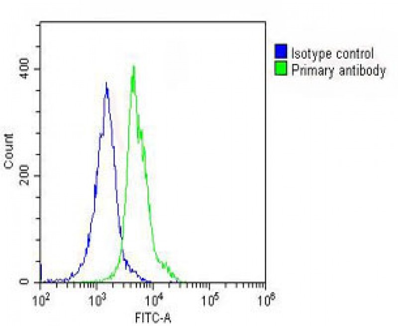
Overlay histogram showing THP-1 cells stained with Antibody (green line). The cells were fixed with 2% paraformaldehyde (10 min) and then permeabilized with 90% methanol for 10 min. The cells were then icubated in 2% bovine serum albumin to block non-specific protein-protein interactions followed by the antibody (1:25 dilution) for 60 min at 37ºC. The secondary antibody used was Goat-Anti-Rabbit IApplication
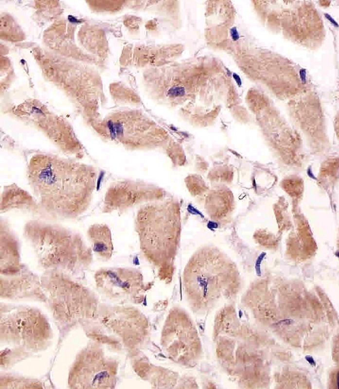
Antibody staining VLDLR in human heart tissue sections by Immunohistochemistry (IHC-P - paraformaldehyde-fixed, paraffin-embedded sections).Application
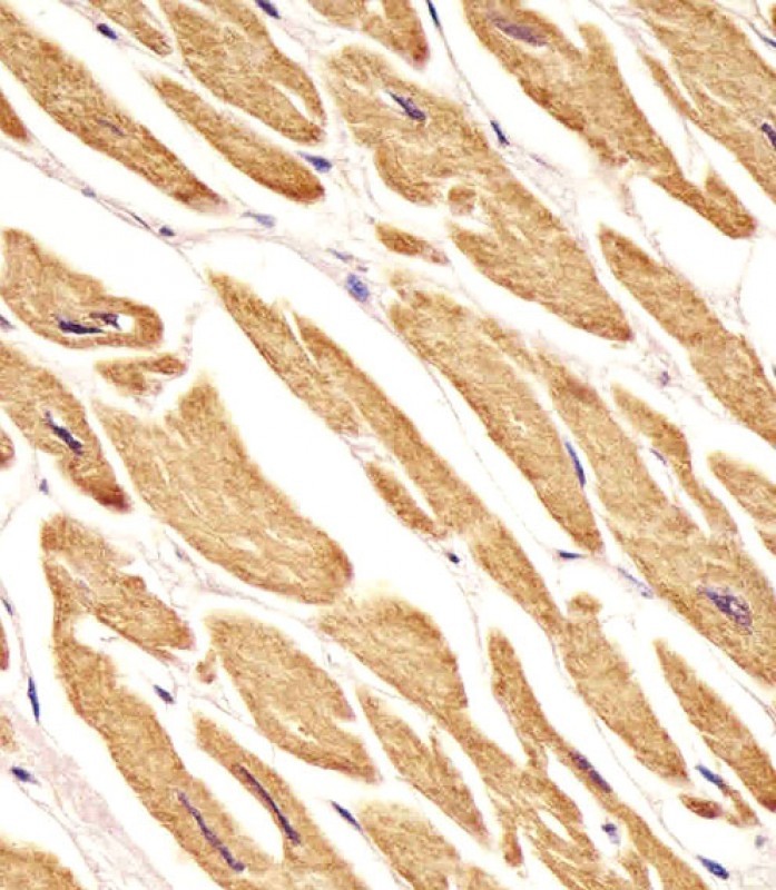
Antibody staining VLDLR in human heart tissue sections by Immunohistochemistry (IHC-P - paraformaldehyde-fixed, paraffin-embedded sections).Application
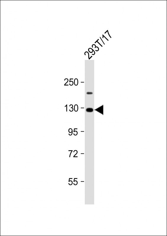
Western Blot at 1:2000 dilution + 293T/17 whole cell lysate Lysates/proteins at 20 ug per lane.Application
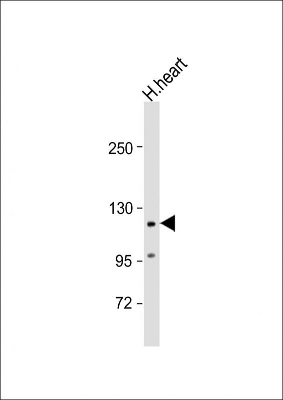
Western Blot at 1:2000 dilution + human heart lysate Lysates/proteins at 20 ug per lane.Application
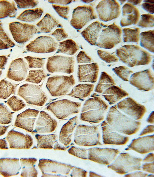
VLDLR Antibody immunohistochemistry analysis in formalin fixed and paraffin embedded human skeletal muscle followed by peroxidase conjugation of the secondary antibody and DAB staining.| Product Name | VLDLR Antibody |
|---|---|
| Antibody Type | Primary Antibodies |
| Product description | The low density lipoprotein receptor (LDLR) gene family consists of cell surface proteins involved in receptor-mediated endocytosis of specific ligands. This gene encodes a lipoprotein receptor that is a member of the LDLR family and plays important roles in VLDL-triglyceride metabolism and the reelin signaling pathway. Mutations in this gene cause VLDLR-associated cerebellar hypoplasia. Alternative splicing generates multiple transcript variants encoding distinct isoforms for this gene. [provided by RefSeq].1) Sakai, K., et al. Brain Res. 1276, 11-21 (2009) |
| Immunogen | This VLDLR antibody is generated from rabbits immunized with a KLH conjugated synthetic peptide between 484-510 amino acids from the Central region of human VLDLR. |
| Clonality | Polyclonal |
|---|---|
| Isotype | Ig |
| Host Species | Rabbit |
| Tested Applications | FACS,IHC-P,WB |
| For FACS starting dilution is: 1:25 For IHC-P starting dilution is: 1:25 For WB starting dilution is: 1:2000 | |
| Species Reactivity | Human |
| Concentration | 1mg/ml |
| Purification | Affinity purified |
| Gene Symbol | VLDLR |
|---|---|
| Alternative Names | Very low-density lipoprotein receptor VLDL receptor VLDL-R VLDLR |
| Molecular Weight(MW) | 96 kDa |
| Sequence Similarities | Predicted species reactivity based on immunogen sequence: Rb |
Application
Overlay histogram showing THP-1 cells stained with Antibody (green line). The cells were fixed with 2% paraformaldehyde (10 min) and then permeabilized with 90% methanol for 10 min. The cells were then icubated in 2% bovine serum albumin to block non-specific protein-protein interactions followed by the antibody (1:25 dilution) for 60 min at 37ºC. The secondary antibody used was Goat-Anti-Rabbit I
Application
Antibody staining VLDLR in human heart tissue sections by Immunohistochemistry (IHC-P - paraformaldehyde-fixed, paraffin-embedded sections).
Application
Antibody staining VLDLR in human heart tissue sections by Immunohistochemistry (IHC-P - paraformaldehyde-fixed, paraffin-embedded sections).
Application
Western Blot at 1:2000 dilution + 293T/17 whole cell lysate Lysates/proteins at 20 ug per lane.
Application
Western Blot at 1:2000 dilution + human heart lysate Lysates/proteins at 20 ug per lane.
Application
VLDLR Antibody immunohistochemistry analysis in formalin fixed and paraffin embedded human skeletal muscle followed by peroxidase conjugation of the secondary antibody and DAB staining.| Application Notes | For FACS starting dilution is: 1:25 For IHC-P starting dilution is: 1:25 For WB starting dilution is: 1:2000 |
|---|
| Form | Liquid |
|---|---|
| Storage Instructions | Store at 4˚C for three months and -20˚C, stable for up to one year. As with all antibodies care should be taken to avoid repeated freeze thaw cycles. Antibodies should not be exposed to prolonged high temperatures. |
| Storage Buffer | Supplied in PBS with 0.09% (W/V) sodium azide. |


 Data sheet for OM294963