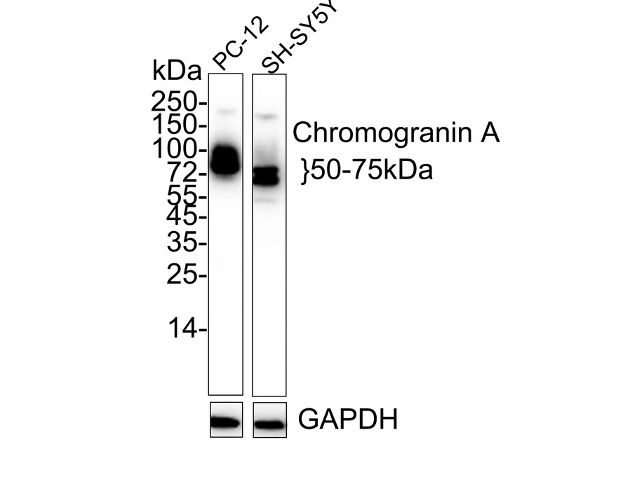
WB
Western blot analysis of Chromogranin A on different lysates with Rabbit anti-Chromogranin A antibody at 1/1,000 dilution. Lane 1: PC-12 cell lysate, Lane 1: SH-SY5Y cell lysate, Lysates/proteins at 20 µg/Lane. Exposure time: 10 seconds; 4-20% SDS-PAGE gel. Proteins were transferred to a PVDF membrane and blocked with 5% NFDM/TBST for 1 hour at room temperature. The primary antibody at 1/1,000 dilution was used in 5% NFDM/TBST at 4℃ overnight. Goat Anti-Rabbit IgG - HRP Secondary Antibody at 1/50,000 dilution was used for 1 hour at room temperature.IHC
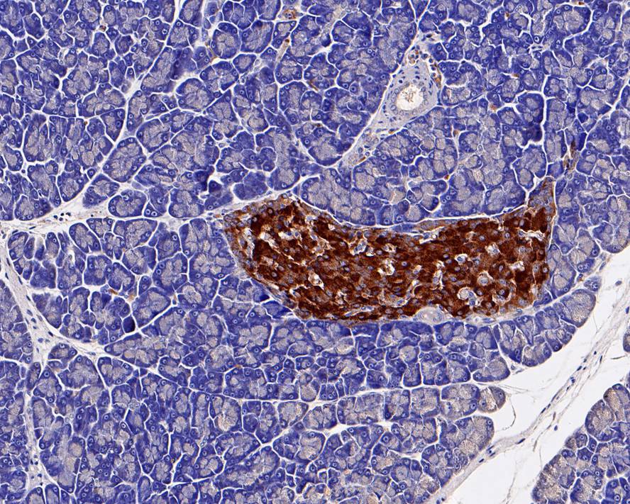
Immunohistochemical analysis of paraffin-embedded rat pancreas tissue with Rabbit anti-Chromogranin A antibody at 1/1,000 dilution. The section was pre-treated using heat mediated antigen retrieval with Tris-EDTA buffer (pH 9.0) for 20 minutes. The tissues were blocked in 1% BSA for 20 minutes at room temperature, washed with ddH2O and PBS, and then probed with the primary antibody at 1/1,000 dilution for 1 hour at room temperature. The detection was performed using an HRP conjugated compact polymer system. DAB was used as the chromogen. Tissues were counterstained with hematoxylin and mounted with DPX.ICC/IF
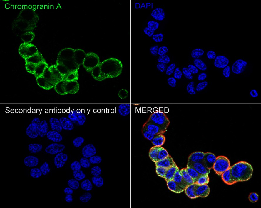
Immunocytochemistry analysis of PC-12 cells labeling Chromogranin A with Rabbit anti-Chromogranin A antibody at 1/100 dilution. Cells were fixed in 4% paraformaldehyde for 20 minutes at room temperature, permeabilized with 0.1% Triton X-100 in PBS for 5 minutes at room temperature, then blocked with 1% BSA in 10% negative goat serum for 1 hour at room temperature. Cells were then incubated with Rabbit anti-Chromogranin A antibody at 1/100 dilution in 1% BSA in PBST overnight at 4 ℃. Goat Anti-Rabbit IgG H&L (iFluor™ 488) was used as the secondary antibody at 1/1,000 dilution. PBS instead of the primary antibody was used as the secondary antibody only control. Nuclear DNA was labelled in blue with DAPI. Beta tubulin (red) was stained at 1/100 dilution overnight at +4℃. Goat Anti-Mouse IgG H&L (iFluor™ 594) was used as the secondary antibody at 1/1,000 dilution.IF-P
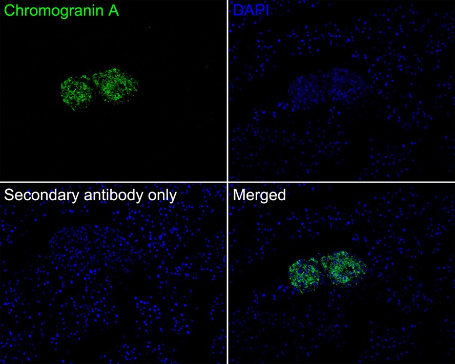
Immunofluorescence analysis of paraffin-embedded mouse pancreas tissue labeling Chromogranin A with Rabbit anti-Chromogranin A antibody at 1/200 dilution. The section was pre-treated using heat mediated antigen retrieval with Tris-EDTA buffer (pH 9.0) for 20 minutes. The tissues were blocked in 10% negative goat serum for 1 hour at room temperature, washed with PBS, and then probed with the primary antibody (green) at 1/200 dilution overnight at 4 ℃, washed with PBS. Goat Anti-Rabbit IgG H&L (iFluor™ 488) was used as the secondary antibody at 1/1,000 dilution. Nuclei were counterstained with DAPI (blue).FC
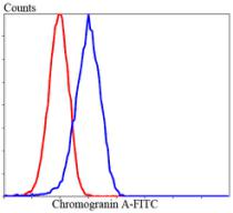
Flow cytometric analysis of Lovo cells with chromogranin A antibody at 1/100 dilution (blue) compared with an unlabelled control (cells without incubation with primary antibody; red). Goat anti rabbit IgG (FITC) was used as the secondary antibody.| Product Name | Chromogranin A Rabbit Polyclonal Antibody |
|---|---|
| Antibody Type | Primary Antibodies |
| Product description | Chromogranins (secretogranins) are acidic glycoproteins that localize within secretory granules of endocrine, neuroendocrine and neuronal tissue. Family members include chromogranin A (Chr-A), chromogranin B (Chr-B, also known as secretogranin I) chromogranin C (also known as secretogranin II or Sg II), secretogranin III (Sg III or SCG3). High levels of Chr-A expression is a characteristic of neuroendocrine tumors. Pancreastatin is a peptide derived from Chr-A which inhibits insulin secretion, exocrine pancreatic secretion and gastric acid secretion. Pancreastatin exists as two forms; the major form is expressed in stomach and colon extracts. In neuroendocrine cells the level Sg II has been shown to increase four-fold in response to histamine, while levels of Chr-A and Chr-B showed little or no increase. Sg III is an acidic secretory protein expressed in neuronal and endocrine cells. In the anterior lobe of the rat pituitary gland, Sg III is present in mammotropes and thyrotropes, moderately in gonadotropes and corticotropes, though not in somatotropes. Sg III and carboxypeptidase E (CPE) bind specifically to cholesterol-rich secretory granule (SG) membranes. |
| Immunogen | Synthetic peptide corresponding to C-terminal of Mouse Chromogranin A aa 408-457 / 457 (ID: P10645). |
| Clonality | polyclonal |
|---|---|
| Isotype | IgG |
| Host Species | Rabbit |
| Tested Applications | FCICC/IFIF-PIHCWB |
| WB:1:2000 IHC:1:200-1:1000 ICC/IF:1:100 IF-P:1:200 FC:1:100-1:200 |
|
| Species Reactivity | HumanMouseRat |
| Concentration | 1mg/ml |
| Purification | Protein A |
| Gene Symbol | CHGA |
|---|---|
| Gene Synonyms | CGA PHE5 PHES |
| Gene Full Name | chromogranin A |
| Alternative Names | beta Granin antibody betagranin (N-terminal fragment of chromogranin A) antibody catestatin antibody CgA antibody CHG A antibody Chga antibody chromofungin antibody Chromogranin A parathyroid secretory protein 1 antibody Chromogranin A precursor antibody ChromograninA antibody CMGA_HUMAN antibody ER-37 antibody Pancreastatin antibody Parastatin antibody Parathyroid secretory protein 1 antibody Pituitary secretory protein I antibody Secretory protein I antibody SP I antibody SP-I antibody SP1 antibody SPI antibody Vasostatin antibody Vasostatin I antibody Vasostatin II antibody |
| Molecular Weight(MW) | 50-75kDa |
| Cellular Localization | Cytoplasmic vesicle, Secreted. |
| SwissProt ID | P10645 |
|---|
WB
Western blot analysis of Chromogranin A on different lysates with Rabbit anti-Chromogranin A antibody at 1/1,000 dilution. Lane 1: PC-12 cell lysate, Lane 1: SH-SY5Y cell lysate, Lysates/proteins at 20 µg/Lane. Exposure time: 10 seconds; 4-20% SDS-PAGE gel. Proteins were transferred to a PVDF membrane and blocked with 5% NFDM/TBST for 1 hour at room temperature. The primary antibody at 1/1,000 dilution was used in 5% NFDM/TBST at 4℃ overnight. Goat Anti-Rabbit IgG - HRP Secondary Antibody at 1/50,000 dilution was used for 1 hour at room temperature.
IHC
Immunohistochemical analysis of paraffin-embedded rat pancreas tissue with Rabbit anti-Chromogranin A antibody at 1/1,000 dilution. The section was pre-treated using heat mediated antigen retrieval with Tris-EDTA buffer (pH 9.0) for 20 minutes. The tissues were blocked in 1% BSA for 20 minutes at room temperature, washed with ddH2O and PBS, and then probed with the primary antibody at 1/1,000 dilution for 1 hour at room temperature. The detection was performed using an HRP conjugated compact polymer system. DAB was used as the chromogen. Tissues were counterstained with hematoxylin and mounted with DPX.
ICC/IF
Immunocytochemistry analysis of PC-12 cells labeling Chromogranin A with Rabbit anti-Chromogranin A antibody at 1/100 dilution. Cells were fixed in 4% paraformaldehyde for 20 minutes at room temperature, permeabilized with 0.1% Triton X-100 in PBS for 5 minutes at room temperature, then blocked with 1% BSA in 10% negative goat serum for 1 hour at room temperature. Cells were then incubated with Rabbit anti-Chromogranin A antibody at 1/100 dilution in 1% BSA in PBST overnight at 4 ℃. Goat Anti-Rabbit IgG H&L (iFluor™ 488) was used as the secondary antibody at 1/1,000 dilution. PBS instead of the primary antibody was used as the secondary antibody only control. Nuclear DNA was labelled in blue with DAPI. Beta tubulin (red) was stained at 1/100 dilution overnight at +4℃. Goat Anti-Mouse IgG H&L (iFluor™ 594) was used as the secondary antibody at 1/1,000 dilution.
IF-P
Immunofluorescence analysis of paraffin-embedded mouse pancreas tissue labeling Chromogranin A with Rabbit anti-Chromogranin A antibody at 1/200 dilution. The section was pre-treated using heat mediated antigen retrieval with Tris-EDTA buffer (pH 9.0) for 20 minutes. The tissues were blocked in 10% negative goat serum for 1 hour at room temperature, washed with PBS, and then probed with the primary antibody (green) at 1/200 dilution overnight at 4 ℃, washed with PBS. Goat Anti-Rabbit IgG H&L (iFluor™ 488) was used as the secondary antibody at 1/1,000 dilution. Nuclei were counterstained with DAPI (blue).
FC
Flow cytometric analysis of Lovo cells with chromogranin A antibody at 1/100 dilution (blue) compared with an unlabelled control (cells without incubation with primary antibody; red). Goat anti rabbit IgG (FITC) was used as the secondary antibody.| Application Notes | WB:1:2000 IHC:1:200-1:1000 ICC/IF:1:100 IF-P:1:200 FC:1:100-1:200 |
|---|
| Form | Liquid |
|---|---|
| Storage Instructions | Store at +4℃ after thawing. Aliquot store at -20℃ or -80℃. Avoid repeated freeze / thaw cycles. |
| Storage Buffer | 1*TBS (pH7.4), 1%BSA, 40%Glycerol. Preservative: 0.05% Sodium Azide. |


 Data sheet for OM637832