



WB
Western blot analysis using GLUT1 antibody against A431 (1) cell lysate.IHC
Immunohistochemical analysis of paraffin-embedded Human lung cancer tissues using GLUT1 antibody with DAB staining.IHC
Immunohistochemical analysis of paraffin-embedded Human breast cancer tissues using GLUT1 antibody with DAB staining.IHC
Immunohistochemical analysis of paraffin-embedded Human lung kidney tissues using GLUT1 antibody with DAB staining.IHC
Immunohistochemical analysis of paraffin-embedded Human ovarian cancer tissues using GLUT1 antibody with DAB staining.IHC
Immunohistochemical analysis of paraffin-embedded Human renal carcinoma tissues using GLUT1 antibody with DAB staining.WB
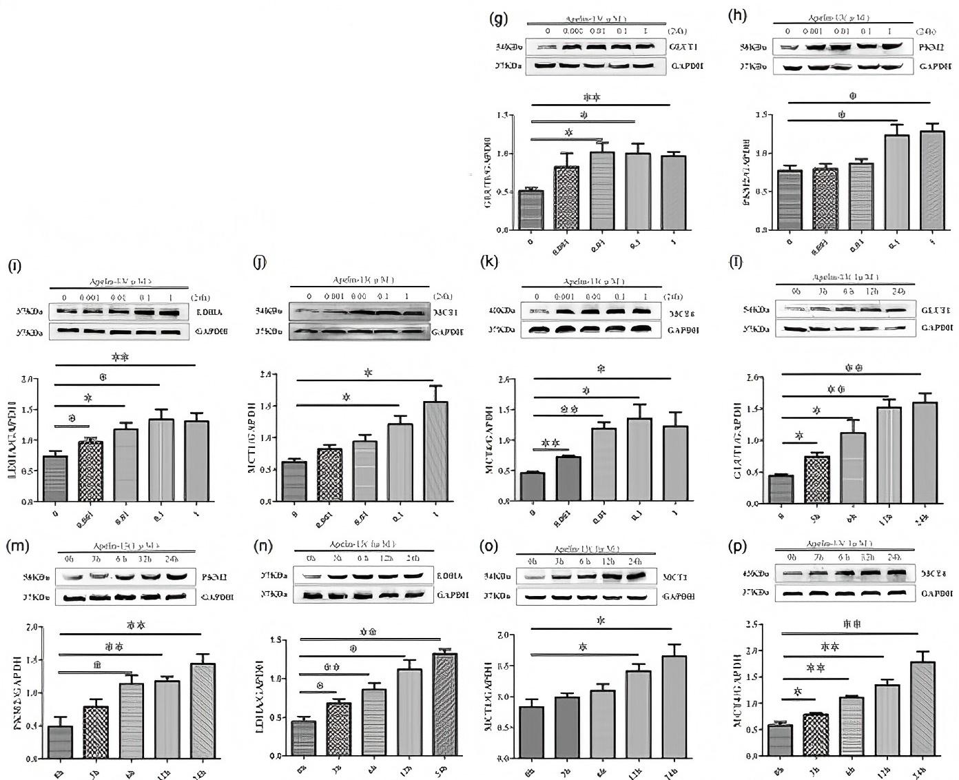
(g–p) Results from western blot analysis showed that apelin-13 promoted the expression of GLUT1, PKM2, LDHA, MCT1, and MCT4 in dose- and time-dependent manners for 24 hr (data were presented as mean ± SEM, n = 3–4; *p < 0.05, **p < 0.01, and ***p < 0.001 vs. control).WB
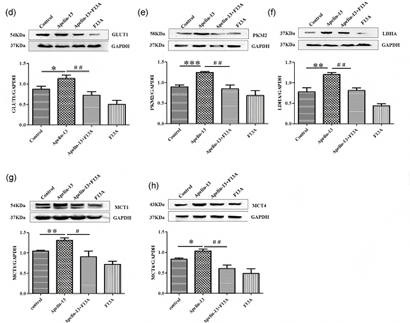
(d–h) Western blot analysis showed that apelin-13 promoted the expression of GLUT1, PKM2, LDHA, MCT1, and MCT4, however, F13A reversed apelin-13-induced GLUT1, PKM2, LDHA, MCT1, and MCT4 expression (data were presented as mean ± SEM, n = 4–5; *p < 0.05, **p < 0.01, and ***p < 0.001 vs.WB
Western blot analysis using GLUT1 antibody against Rat stomach(1) tissue.| Product Name | Anti-GLUT1 antibody-poly-HRP |
|---|---|
| Antibody Type | Primary Antibodies |
| Immunogen | Polypeptide |
| Conjugation | poly-HRP |
| Clonality | Polyclonal |
|---|---|
| Isotype | IgG |
| Host Species | Rabbit |
| Tested Applications | WBIHC |
| WB:1:200-1:2000 IHC:1:100-1:1000 |
|
| Species Reactivity | HumanMouseRat |
| Concentration | 1mg/ml |
| Purification | Protein A |
| Gene Symbol | SLC2A1 |
|---|---|
| Gene Synonyms | GTR1 SLC2A1 CSE PED DYT9 GLUT DYT17 DYT18 EIG12 GLUT1 HTLVR GLUT-1 SDCHCN GLUT1DS |
| Gene Full Name | solute carrier family 2 member 1 |
| Gene Summary | This gene encodes a major glucose transporter in the mammalian blood-brain barrier. The encoded protein is found primarily in the cell membrane and on the cell surface, where it can also function as a receptor for human T-cell leukemia virus (HTLV) I and II. Mutations in this gene have been found in a family with paroxysmal exertion-induced dyskinesia. [provided by RefSeq, Apr 2013] |
| Molecular Weight(MW) | 54 KDa |
| Source | Rabbit |
| Cellular Localization | Cell membrane. Melanosome |
| SwissProt ID | P11166 |
|---|
WB
Western blot analysis using GLUT1 antibody against A431 (1) cell lysate.
IHC
Immunohistochemical analysis of paraffin-embedded Human lung cancer tissues using GLUT1 antibody with DAB staining.
IHC
Immunohistochemical analysis of paraffin-embedded Human breast cancer tissues using GLUT1 antibody with DAB staining.
IHC
Immunohistochemical analysis of paraffin-embedded Human lung kidney tissues using GLUT1 antibody with DAB staining.
IHC
Immunohistochemical analysis of paraffin-embedded Human ovarian cancer tissues using GLUT1 antibody with DAB staining.
IHC
Immunohistochemical analysis of paraffin-embedded Human renal carcinoma tissues using GLUT1 antibody with DAB staining.
WB
(g–p) Results from western blot analysis showed that apelin-13 promoted the expression of GLUT1, PKM2, LDHA, MCT1, and MCT4 in dose- and time-dependent manners for 24 hr (data were presented as mean ± SEM, n = 3–4; *p < 0.05, **p < 0.01, and ***p < 0.001 vs. control).
WB
(d–h) Western blot analysis showed that apelin-13 promoted the expression of GLUT1, PKM2, LDHA, MCT1, and MCT4, however, F13A reversed apelin-13-induced GLUT1, PKM2, LDHA, MCT1, and MCT4 expression (data were presented as mean ± SEM, n = 4–5; *p < 0.05, **p < 0.01, and ***p < 0.001 vs.
WB
Western blot analysis using GLUT1 antibody against Rat stomach(1) tissue.| Application Notes | WB:1:200-1:2000 IHC:1:100-1:1000 |
|---|
| Form | Liquid |
|---|---|
| Storage Instructions | Shipped at 4°C. Store at +4°C short term (1-2 weeks). Store at -20°C long term. Avoid freeze / thaw cycle. |
| Storage Buffer | Purified antibody in PBS with 0.05% sodium azide. |


