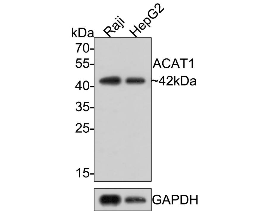
WB
Western blot analysis of ACAT-1 on different lysates with Rabbit anti-ACAT-1 antibody at 1/500 dilution. Lane 1: Raji cell lysate Lane 2: HepG2 cell lysate Lysates/proteins at 10 µg/Lane. Predicted band size: 45 kDa Observed band size: 42 kDa Exposure time: 4 minutes; 12% SDS-PAGE gel. Proteins were transferred to a PVDF membrane and blocked with 5% NFDM/TBST for 1 hour at room temperature. The primary antibody at 1/500 dilution was used in 5% NFDM/TBST at room temperature for 2 hours. Goat Anti-Rabbit IgG - HRP Secondary Antibody at 1:200,000 dilution was used for 1 hour at room temperature.IHC
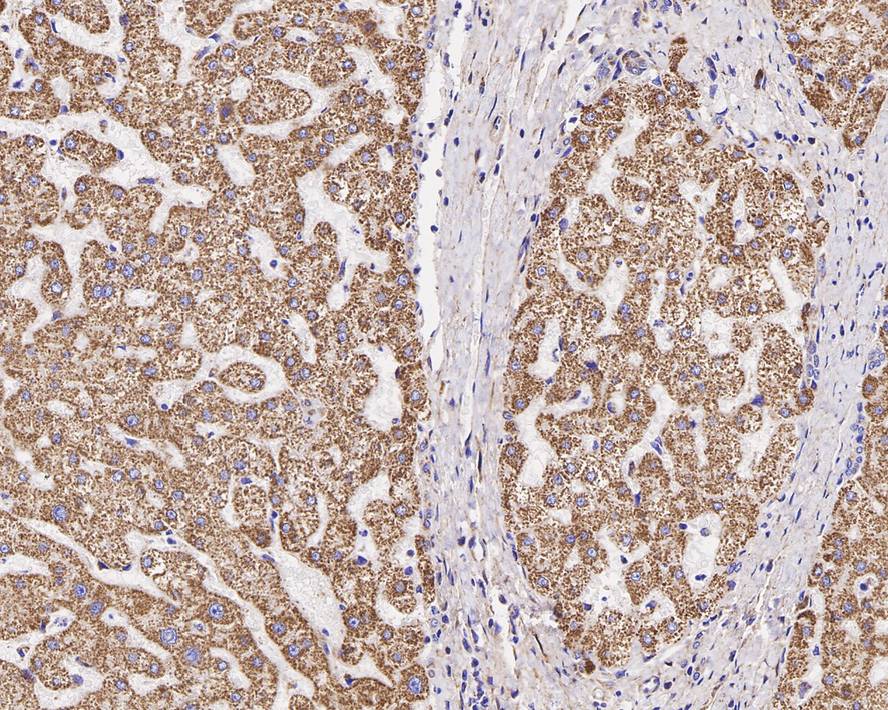
Immunohistochemical analysis of paraffin-embedded human liver tissue with Rabbit anti-ACAT-1 antibody at 1/200 dilution. The section was pre-treated using heat mediated antigen retrieval with Tris-EDTA buffer (pH 9.0) for 20 minutes. The tissues were blocked in 1% BSA for 20 minutes at room temperature, washed with ddH2O and PBS, and then probed with the primary antibody at 1/200 dilution for 1 hour at room temperature. The detection was performed using an HRP conjugated compact polymer system. DAB was used as the chromogen. Tissues were counterstained with hematoxylin and mounted with DPX.ICC/IF
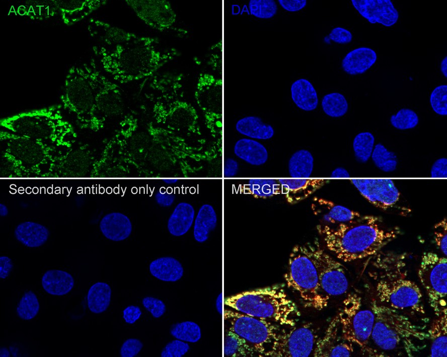
Immunocytochemistry analysis of HepG2 cells labeling ACAT-1 with Rabbit anti-ACAT-1 antibody at 1/250 dilution. Cells were fixed in 4% paraformaldehyde for 20 minutes at room temperature, permeabilized with 0.1% Triton X-100 in PBS for 5 minutes at room temperature, then blocked with 1% BSA in 10% negative goat serum for 1 hour at room temperature. Cells were then incubated with Rabbit anti-ACAT-1 antibody at 1/250 dilution in 1% BSA in PBST overnight at 4 ℃. Goat Anti-Rabbit IgG H&L (iFluor™ 488) was used as the secondary antibody at 1/1,000 dilution. PBS instead of the primary antibody was used as the secondary antibody only control. Counterstained with Mitotracker. Nuclear DNA was labelled in blue with DAPI.| Product Name | ACAT-1 Recombinant Rabbit Monoclonal Antibody |
|---|---|
| Antibody Type | Primary Antibodies |
| Immunogen | Synthetic peptide within human ACAT1 aa 378-427/427. |
| Clonality | Monoclonal |
|---|---|
| Isotype | IgG |
| Host Species | Rabbit |
| Tested Applications | ICC/IFIHCWB |
| WB:1:500-1:1000 IHC:1:100-1:400 ICC:1:250 |
|
| Species Reactivity | HumanMouseRat |
| Concentration | 1mg/ml |
| Purification | Protein A |
| Gene Symbol | ACAT1 |
|---|---|
| Gene Synonyms | T2 MAT ACAT THIL |
| Gene Full Name | acetyl-CoA acetyltransferase 1 |
| Gene Summary | This gene encodes a mitochondrially localized enzyme that catalyzes the reversible formation of acetoacetyl-CoA from two molecules of acetyl-CoA. Defects in this gene are associated with 3-ketothiolase deficiency, an inborn error of isoleucine catabolism characterized by urinary excretion of 2-methyl-3-hydroxybutyric acid, 2-methylacetoacetic acid, tiglylglycine, and butanone. [provided by RefSeq, Feb 2009] |
| Molecular Weight(MW) | 45kDa |
| Cellular Localization | Mitochondrion. |
WB
Western blot analysis of ACAT-1 on different lysates with Rabbit anti-ACAT-1 antibody at 1/500 dilution. Lane 1: Raji cell lysate Lane 2: HepG2 cell lysate Lysates/proteins at 10 µg/Lane. Predicted band size: 45 kDa Observed band size: 42 kDa Exposure time: 4 minutes; 12% SDS-PAGE gel. Proteins were transferred to a PVDF membrane and blocked with 5% NFDM/TBST for 1 hour at room temperature. The primary antibody at 1/500 dilution was used in 5% NFDM/TBST at room temperature for 2 hours. Goat Anti-Rabbit IgG - HRP Secondary Antibody at 1:200,000 dilution was used for 1 hour at room temperature.
IHC
Immunohistochemical analysis of paraffin-embedded human liver tissue with Rabbit anti-ACAT-1 antibody at 1/200 dilution. The section was pre-treated using heat mediated antigen retrieval with Tris-EDTA buffer (pH 9.0) for 20 minutes. The tissues were blocked in 1% BSA for 20 minutes at room temperature, washed with ddH2O and PBS, and then probed with the primary antibody at 1/200 dilution for 1 hour at room temperature. The detection was performed using an HRP conjugated compact polymer system. DAB was used as the chromogen. Tissues were counterstained with hematoxylin and mounted with DPX.
ICC/IF
Immunocytochemistry analysis of HepG2 cells labeling ACAT-1 with Rabbit anti-ACAT-1 antibody at 1/250 dilution. Cells were fixed in 4% paraformaldehyde for 20 minutes at room temperature, permeabilized with 0.1% Triton X-100 in PBS for 5 minutes at room temperature, then blocked with 1% BSA in 10% negative goat serum for 1 hour at room temperature. Cells were then incubated with Rabbit anti-ACAT-1 antibody at 1/250 dilution in 1% BSA in PBST overnight at 4 ℃. Goat Anti-Rabbit IgG H&L (iFluor™ 488) was used as the secondary antibody at 1/1,000 dilution. PBS instead of the primary antibody was used as the secondary antibody only control. Counterstained with Mitotracker. Nuclear DNA was labelled in blue with DAPI.| Application Notes | WB:1:500-1:1000 IHC:1:100-1:400 ICC:1:250 |
|---|
| Form | Liquid |
|---|---|
| Storage Instructions | Store at +4℃ after thawing. Aliquot store at -20℃. Avoid repeated freeze / thaw cycles. |
| Storage Buffer | 1*TBS (pH7.4), 0.05% BSA, 40% Glycerol. Preservative: 0.05% Sodium Azide. |


 Data sheet for OM643039