IHC
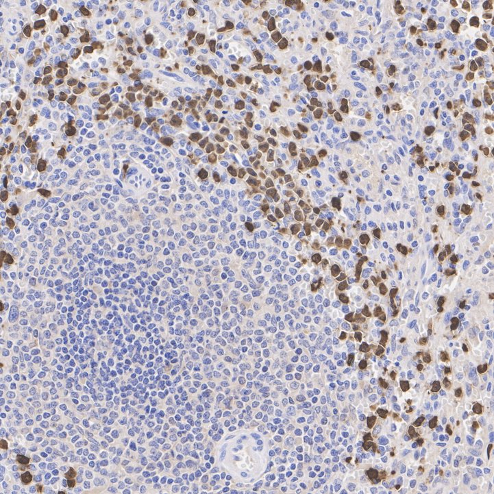
Immunohistochemical analysis of paraffin-embedded human spleen tissue with Rabbit anti-S100A9 antibody at 1/1,000 dilution. The section was pre-treated using heat mediated antigen retrieval with Tris-EDTA buffer (pH 9.0) for 20 minutes. The tissues were blocked in 1% BSA for 20 minutes at room temperature, washed with ddH2O and PBS, and then probed with the primary antibody at 1/1,000 dilution for 1 hour at room temperature. The detection was performed using an HRP conjugated compact polymer system. DAB was used as the chromogen. Tissues were counterstained with hematoxylin and mounted with DPX.mIHC
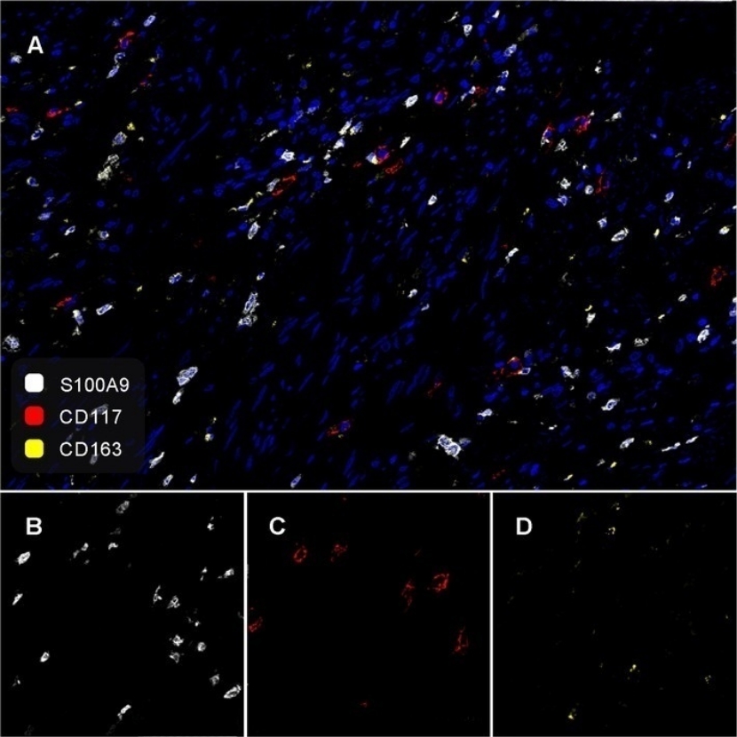
Fluorescence multiplex immunohistochemical analysis of human cervical carcinoma (Formalin/PFA-fixed paraffin-embedded sections). Panel A: the merged image of anti-S100A9 (White), anti-CD117 (Red) and anti-CD163(Yellow) on human cervical carcinoma. HRP Conjugated UltraPolymer Goat Polyclonal Antibody HA1119/HA1120 was used as a secondary antibody. The immunostaining was performed with the Sequential Immuno-staining Kit (IRISKit™MH010101, www.luminiris.cn). The section was incubated in three rounds of staining: in the order of anti-S100A9 (1/1,000 dilution),anti-CD117 (1/1,000 dilution) and anti-CD163 (1/2,000 dilution) for 20 mins at room temperature. Each round was followed by a separate fluorescent tyramide signal amplification system. Heat mediated antigen retrieval with Tris-EDTA buffer (pH 9.0) for 30 mins at 95℃. DAPI (blue) was used as a nuclear counter stain. Image acquisition was performed with Zeiss Observer 7 Inverted Fluorescence Microscope.| Product Name | S100A9 Recombinant Rabbit Monoclonal Antibody |
|---|---|
| Antibody Type | Primary Antibodies |
| Immunogen | Synthetic peptide within Human S100A9 aa 1-42 / 114. |
| Clonality | Monoclonal |
|---|---|
| Isotype | IgG |
| Host Species | Rabbit |
| Tested Applications | IF-PIHC |
| IHC:1:1000 IF-P:1:50-1:200 |
|
| Species Reactivity | Human |
| Concentration | 1mg/ml |
| Purification | Protein A |
| Gene Symbol | S100A9 |
|---|---|
| Gene Synonyms | MIF NIF P14 CAGB CFAG CGLB L1AG LIAG MRP14 60B8AG MAC387 S100-A9 |
| Gene Full Name | S100 calcium binding protein A9 |
| Gene Summary | The protein encoded by this gene is a member of the S100 family of proteins containing 2 EF-hand calcium-binding motifs. S100 proteins are localized in the cytoplasm and/or nucleus of a wide range of cells, and involved in the regulation of a number of cellular processes such as cell cycle progression and differentiation. S100 genes include at least 13 members which are located as a cluster on chromosome 1q21. This protein may function in the inhibition of casein kinase and altered expression of this protein is associated with the disease cystic fibrosis. This antimicrobial protein exhibits antifungal and antibacterial activity. [provided by RefSeq, Nov 2014] |
| Molecular Weight(MW) | 13kDa |
| Cellular Localization | Secreted, Cytoplasm, Cell membrane. |
IHC
Immunohistochemical analysis of paraffin-embedded human spleen tissue with Rabbit anti-S100A9 antibody at 1/1,000 dilution. The section was pre-treated using heat mediated antigen retrieval with Tris-EDTA buffer (pH 9.0) for 20 minutes. The tissues were blocked in 1% BSA for 20 minutes at room temperature, washed with ddH2O and PBS, and then probed with the primary antibody at 1/1,000 dilution for 1 hour at room temperature. The detection was performed using an HRP conjugated compact polymer system. DAB was used as the chromogen. Tissues were counterstained with hematoxylin and mounted with DPX.
mIHC
Fluorescence multiplex immunohistochemical analysis of human cervical carcinoma (Formalin/PFA-fixed paraffin-embedded sections). Panel A: the merged image of anti-S100A9 (White), anti-CD117 (Red) and anti-CD163(Yellow) on human cervical carcinoma. HRP Conjugated UltraPolymer Goat Polyclonal Antibody HA1119/HA1120 was used as a secondary antibody. The immunostaining was performed with the Sequential Immuno-staining Kit (IRISKit™MH010101, www.luminiris.cn). The section was incubated in three rounds of staining: in the order of anti-S100A9 (1/1,000 dilution),anti-CD117 (1/1,000 dilution) and anti-CD163 (1/2,000 dilution) for 20 mins at room temperature. Each round was followed by a separate fluorescent tyramide signal amplification system. Heat mediated antigen retrieval with Tris-EDTA buffer (pH 9.0) for 30 mins at 95℃. DAPI (blue) was used as a nuclear counter stain. Image acquisition was performed with Zeiss Observer 7 Inverted Fluorescence Microscope.| Application Notes | IHC:1:1000 IF-P:1:50-1:200 |
|---|
| Form | Liquid |
|---|---|
| Storage Instructions | Store at +4℃ after thawing. Aliquot store at -20℃ or -80℃. Avoid repeated freeze / thaw cycles. |
| Storage Buffer | 1*TBS (pH7.4), 0.05% BSA, 40% Glycerol. Preservative: 0.05% Sodium Azide. |


 Data sheet for OM643174