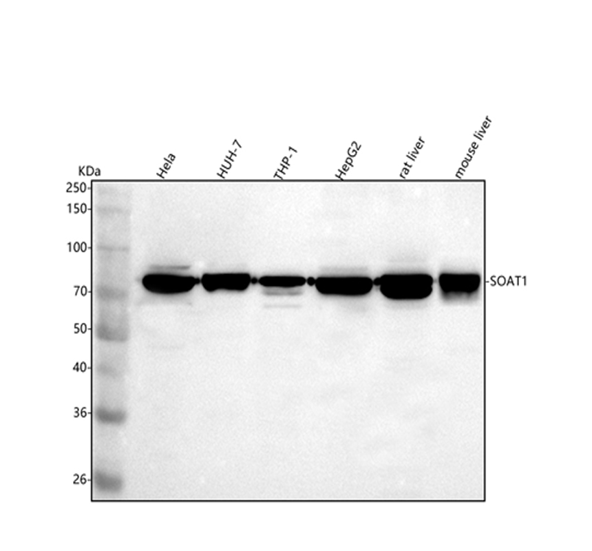
WB
Western blot analysis of SOAT1 using anti-SOAT1 antibody. The sample well of each lane was loaded with 30 ug of sample under reducing conditions. Lane 1: human Hela whole cell lysates, Lane 2: human HUH-7 whole cell lysates, Lane 3: human THP-1 whole cell lysates, Lane 4: human HepG2 whole cell lysates, Lane 5: rat liver tissue lysates, Lane 6: mouse liver tissue lysates. After electrophoresis, proteins were transferred to a membrane. Then the membrane was incubated with rabbit anti-SOAT1 antigen affinity purified polyclonal antibody at a dilution of 1:1000 and probed with a goat anti-rabbit IgG-HRP secondary antibody. The signal is developed using ECL Plus Western Blotting Substrate. A specific band was detected for SOAT1 at approximately 75 kDa. The expected band size for SOAT1 is at 65 kDa.IHC
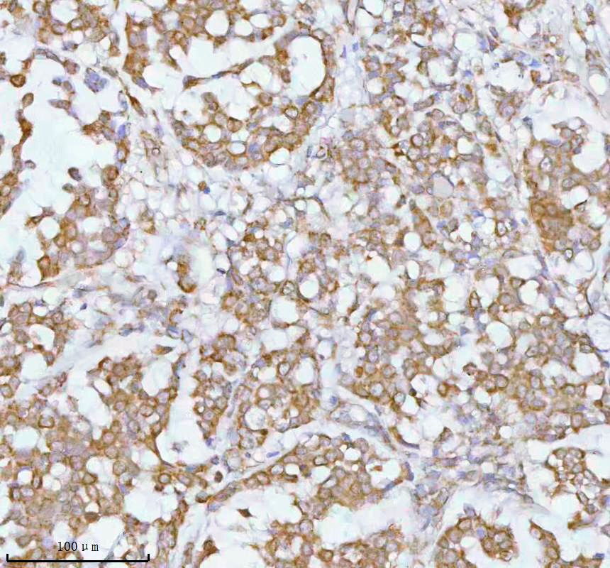
IHC analysis of SOAT1 using anti-SOAT1 antibody. SOAT1 was detected in a paraffin-embedded section of human breast cancer tissue. The tissue section was incubated with rabbit anti-SOAT1 Antibody at a dilution of 1:200 and developed using HRP Conjugated Rabbit IgG Super Vision Assay Kit with DAB as the chromogen.ICC/IF
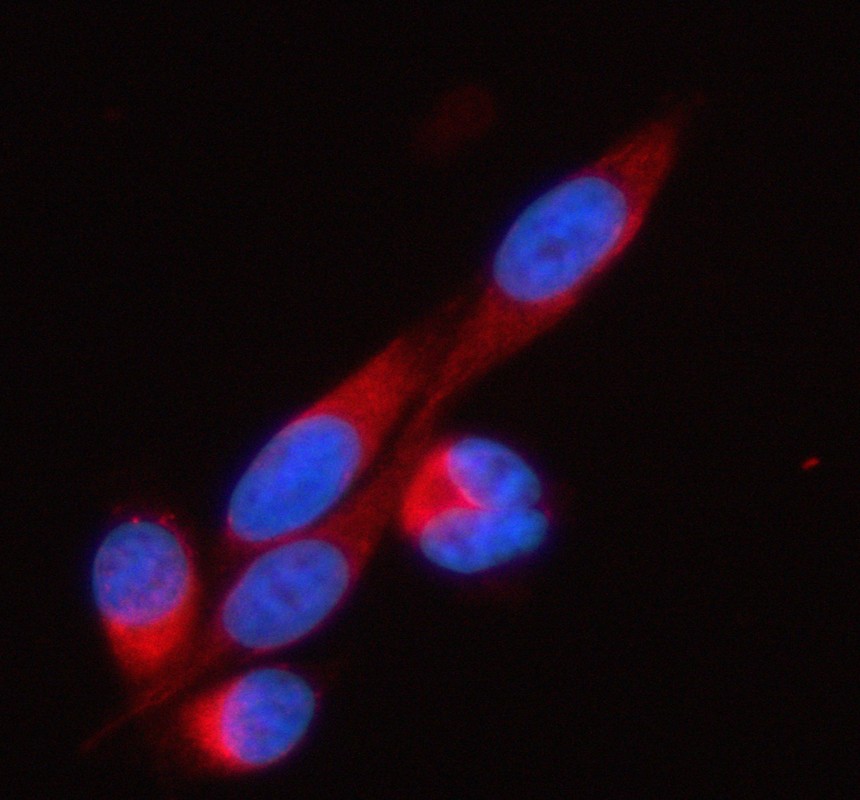
IF analysis of SOAT1 using anti-SOAT1 antibody. SOAT1 was detected in an immunocytochemical section of PC-3 cells. The section was incubated with rabbit anti-SOAT1 Antibody at a dilution of 1:100. Cy3-conjugated Anti-rabbit IgG Secondary Antibody (red)was used as secondary antibody. The section was counterstained with DAPI (Blue).FC
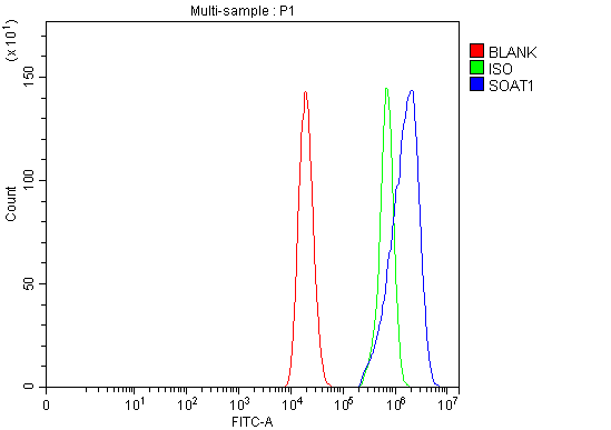
Flow Cytometry analysis of Caco-2 cells using anti-SOAT1 antibody. Overlay histogram showing Caco-2 cells stained with anti-SOAT1 antibody (Blue line). To facilitate intracellular staining, cells were fixed with 4% paraformaldehyde and permeabilized with permeabilization buffer. The cells were blocked with 10% normal goat serum. And then incubated with rabbit anti-SOAT1 Antibody at 1:100 dilution for 30 min at 20°C. DyLight®488 conjugated goat anti-rabbit IgG was used as secondary antibody at 1:100 dilution for 30 minutes at 20°C. Isotype control antibody (Green line) was rabbit IgG at 1:100 dilution used under the same conditions. Unlabelled sample (Red line) was also used as a control.| Product Name | Rabbit polyclonal antibody to SOAT1 |
|---|---|
| Antibody Type | Primary Antibodies |
| Immunogen | E.coli-derived human SOAT1 recombinant protein (Position: R41-Y548). Human SOAT1 shares 88.2% and 87.4% amino acid (aa) sequence identity with mouse and rat SOAT1, respectively. |
| Clonality | Polyclonal |
|---|---|
| Isotype | IgG |
| Host Species | Rabbit |
| Tested Applications | FCICC/IFIHCWB |
| WB:1:500-1:2000 IHC:1:50-1:400 ICC:1:50-1:400 FC:1:50-1:200 |
|
| Species Reactivity | HumanMouseRat |
| Concentration | 0.5mg/ml |
| Purification | Affinity purified |
| Gene Symbol | SOAT1 |
|---|---|
| Gene Synonyms | ACAT SOAT STAT ACACT ACAT1 ACAT-1 |
| Gene Full Name | sterol O-acyltransferase 1 |
| Gene Summary | The protein encoded by this gene belongs to the acyltransferase family. It is located in the endoplasmic reticulum, and catalyzes the formation of fatty acid-cholesterol esters. This gene has been implicated in the formation of beta-amyloid and atherosclerotic plaques by controlling the equilibrium between free cholesterol and cytoplasmic cholesteryl esters. Alternatively spliced transcript variants have been found for this gene. [provided by RefSeq, Nov 2011] |
| Molecular Weight(MW) | 65kDa(Observed MW 75kDa) |
| Cellular Localization | Endoplasmic reticulum membrane. |
WB
Western blot analysis of SOAT1 using anti-SOAT1 antibody. The sample well of each lane was loaded with 30 ug of sample under reducing conditions. Lane 1: human Hela whole cell lysates, Lane 2: human HUH-7 whole cell lysates, Lane 3: human THP-1 whole cell lysates, Lane 4: human HepG2 whole cell lysates, Lane 5: rat liver tissue lysates, Lane 6: mouse liver tissue lysates. After electrophoresis, proteins were transferred to a membrane. Then the membrane was incubated with rabbit anti-SOAT1 antigen affinity purified polyclonal antibody at a dilution of 1:1000 and probed with a goat anti-rabbit IgG-HRP secondary antibody. The signal is developed using ECL Plus Western Blotting Substrate. A specific band was detected for SOAT1 at approximately 75 kDa. The expected band size for SOAT1 is at 65 kDa.
IHC
IHC analysis of SOAT1 using anti-SOAT1 antibody. SOAT1 was detected in a paraffin-embedded section of human breast cancer tissue. The tissue section was incubated with rabbit anti-SOAT1 Antibody at a dilution of 1:200 and developed using HRP Conjugated Rabbit IgG Super Vision Assay Kit with DAB as the chromogen.
ICC/IF
IF analysis of SOAT1 using anti-SOAT1 antibody. SOAT1 was detected in an immunocytochemical section of PC-3 cells. The section was incubated with rabbit anti-SOAT1 Antibody at a dilution of 1:100. Cy3-conjugated Anti-rabbit IgG Secondary Antibody (red)was used as secondary antibody. The section was counterstained with DAPI (Blue).
FC
Flow Cytometry analysis of Caco-2 cells using anti-SOAT1 antibody. Overlay histogram showing Caco-2 cells stained with anti-SOAT1 antibody (Blue line). To facilitate intracellular staining, cells were fixed with 4% paraformaldehyde and permeabilized with permeabilization buffer. The cells were blocked with 10% normal goat serum. And then incubated with rabbit anti-SOAT1 Antibody at 1:100 dilution for 30 min at 20°C. DyLight®488 conjugated goat anti-rabbit IgG was used as secondary antibody at 1:100 dilution for 30 minutes at 20°C. Isotype control antibody (Green line) was rabbit IgG at 1:100 dilution used under the same conditions. Unlabelled sample (Red line) was also used as a control.| Application Notes | WB:1:500-1:2000 IHC:1:50-1:400 ICC:1:50-1:400 FC:1:50-1:200 |
|---|
| Form | Liquid |
|---|---|
| Storage Instructions | 12 months from date of receipt, -20℃ as supplied. 6 months 2 to 8℃ after reconstitution. Avoid repeated freezing and thawing. |
| Storage Buffer | 500 ug/ml antibody with PBS, 0.02% NaN3, 1 mg/ml BSA and 50% glycerol. |


 Data sheet for OM643539