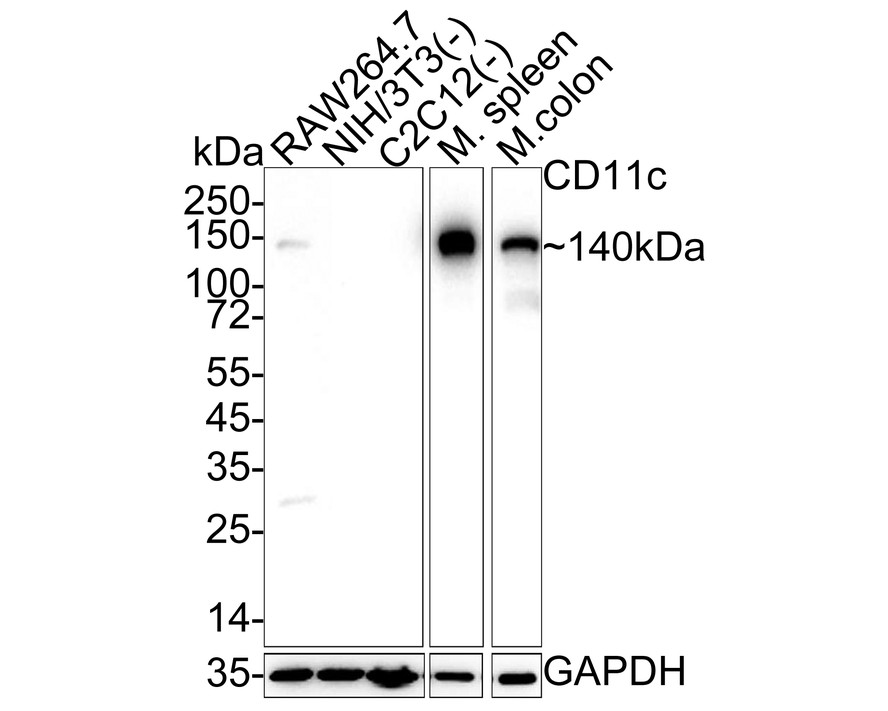
WB
Western blot analysis of CD11c on different lysates with Rabbit anti-CD11c antibody at 1/1,000 dilution. Lane 1: RAW264.7 cell lysate (20 µg/Lane) Lane 2:NIH/3T3 cell lysate (negative) (20 µg/Lane) Lane 3: C2C12 cell lysate (negative) (20 µg/Lane) Lane 4: Mouse spleen tissue lysate (30 µg/Lane) Lane 5: Mouse colon tissue lysate (30 µg/Lane) Exposure time: Lane1-3: 3 minutes ; Lane 4-5: 25 seconds; 4-20% SDS-PAGE gel. Proteins were transferred to a PVDF membrane and blocked with 5% NFDM/TBST for 1 hour at room temperature. The primary antibody at 1/1,000 dilution was used in 5% NFDM/TBST at 4℃ overnight. Goat Anti-Rabbit IgG - HRP Secondary Antibody at 1/50,000 dilution was used for 1 hour at room temperature.IHC
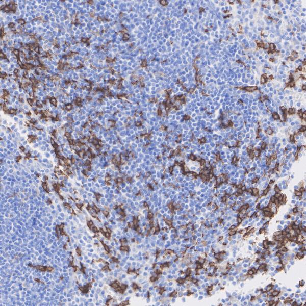
Immunohistochemical analysis of paraffin-embedded mouse spleen tissue with Rabbit anti-CD11c antibody at 1/1,000 dilution. The section was pre-treated using heat mediated antigen retrieval with Tris-EDTA buffer (pH 9.0) for 20 minutes. The tissues were blocked in 1% BSA for 20 minutes at room temperature, washed with ddH2O and PBS, and then probed with the primary antibody at 1/1,000 dilution for 1 hour at room temperature. The detection was performed using an HRP conjugated compact polymer system. DAB was used as the chromogen. Tissues were counterstained with hematoxylin and mounted with DPX.IF-F
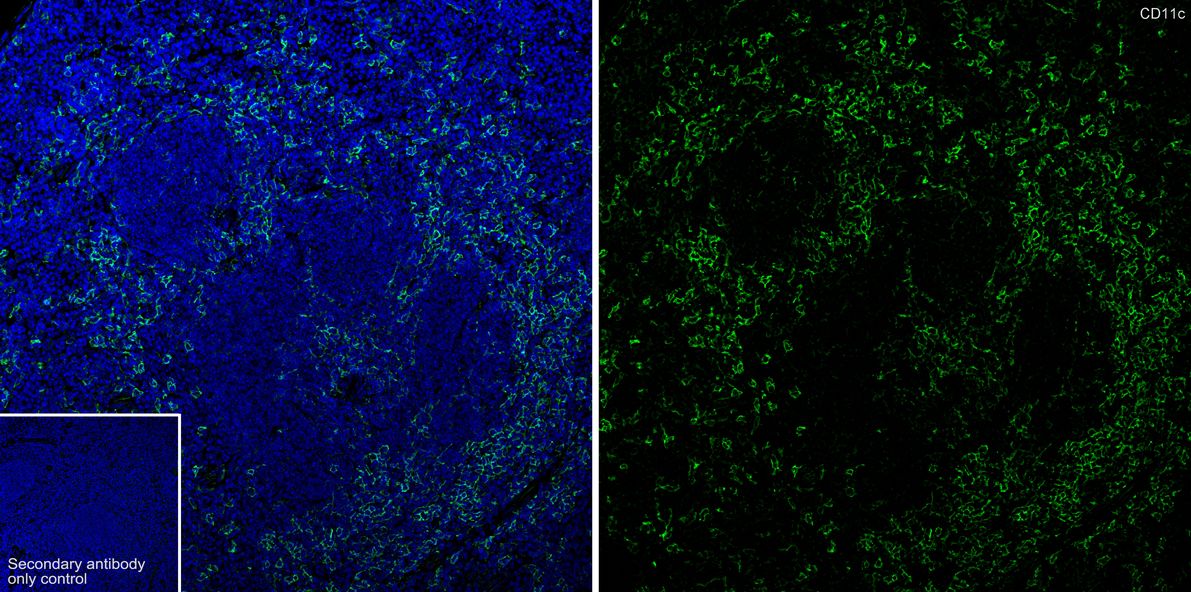
Immunofluorescence analysis of frozen mouse spleen tissue with Rabbit anti-CD11c antibody at 1/200 dilution. The section was pre-treated using heat mediated antigen retrieval with sodium citrate buffer (pH 6.0) for about 2 minutes in microwave oven. The tissues were blocked in 10% negative goat serum for 1 hour at room temperature, washed with PBS, and then probed with the primary antibody (green) at 1/200 dilution overnight at 4 ℃, washed with PBS. Goat Anti-Rabbit IgG H&L (iFluor™ 488) was used as the secondary antibody at 1/1,000 dilution. Nuclei were counterstained with DAPI (blue).| Product Name | CD11c Recombinant Rabbit Monoclonal Antibody |
|---|---|
| Antibody Type | Primary Antibodies |
| Immunogen | Recombinant protein within mouse CD11c aa 616-936 / 1,169. |
| Clonality | Monoclonal |
|---|---|
| Isotype | IgG |
| Host Species | Rabbit |
| Tested Applications | IF-FIHCWB |
| WB:1:1000 IHC:1:1000 IF-F:1:200 |
|
| Species Reactivity | Mouse |
| Concentration | 1mg/ml |
| Purification | Protein A |
| Gene Symbol | Itgax |
|---|---|
| Gene Synonyms | CD11C SLEB6 |
| Gene Full Name | integrin subunit alpha X |
| Gene Summary | This gene encodes the integrin alpha X chain protein. Integrins are heterodimeric integral membrane proteins composed of an alpha chain and a beta chain. This protein combines with the beta 2 chain (ITGB2) to form a leukocyte-specific integrin referred to as inactivated-C3b (iC3b) receptor 4 (CR4). The alpha X beta 2 complex seems to overlap the properties of the alpha M beta 2 integrin in the adherence of neutrophils and monocytes to stimulated endothelium cells, and in the phagocytosis of complement coated particles. Two transcript variants encoding different isoforms have been found for this gene. [provided by RefSeq, Nov 2013] |
| Molecular Weight(MW) | 129 kDa(Observed band size: 140kDa) |
| Cellular Localization | Membrane |
WB
Western blot analysis of CD11c on different lysates with Rabbit anti-CD11c antibody at 1/1,000 dilution. Lane 1: RAW264.7 cell lysate (20 µg/Lane) Lane 2:NIH/3T3 cell lysate (negative) (20 µg/Lane) Lane 3: C2C12 cell lysate (negative) (20 µg/Lane) Lane 4: Mouse spleen tissue lysate (30 µg/Lane) Lane 5: Mouse colon tissue lysate (30 µg/Lane) Exposure time: Lane1-3: 3 minutes ; Lane 4-5: 25 seconds; 4-20% SDS-PAGE gel. Proteins were transferred to a PVDF membrane and blocked with 5% NFDM/TBST for 1 hour at room temperature. The primary antibody at 1/1,000 dilution was used in 5% NFDM/TBST at 4℃ overnight. Goat Anti-Rabbit IgG - HRP Secondary Antibody at 1/50,000 dilution was used for 1 hour at room temperature.
IHC
Immunohistochemical analysis of paraffin-embedded mouse spleen tissue with Rabbit anti-CD11c antibody at 1/1,000 dilution. The section was pre-treated using heat mediated antigen retrieval with Tris-EDTA buffer (pH 9.0) for 20 minutes. The tissues were blocked in 1% BSA for 20 minutes at room temperature, washed with ddH2O and PBS, and then probed with the primary antibody at 1/1,000 dilution for 1 hour at room temperature. The detection was performed using an HRP conjugated compact polymer system. DAB was used as the chromogen. Tissues were counterstained with hematoxylin and mounted with DPX.
IF-F
Immunofluorescence analysis of frozen mouse spleen tissue with Rabbit anti-CD11c antibody at 1/200 dilution. The section was pre-treated using heat mediated antigen retrieval with sodium citrate buffer (pH 6.0) for about 2 minutes in microwave oven. The tissues were blocked in 10% negative goat serum for 1 hour at room temperature, washed with PBS, and then probed with the primary antibody (green) at 1/200 dilution overnight at 4 ℃, washed with PBS. Goat Anti-Rabbit IgG H&L (iFluor™ 488) was used as the secondary antibody at 1/1,000 dilution. Nuclei were counterstained with DAPI (blue).| Application Notes | WB:1:1000 IHC:1:1000 IF-F:1:200 |
|---|
| Form | Liquid |
|---|---|
| Storage Instructions | Store at +4℃ after thawing. Aliquot store at -20℃. Avoid repeated freeze / thaw cycles. |
| Storage Buffer | PBS (pH7.4), 0.1% BSA, 40% Glycerol. Preservative: 0.05% Sodium Azide. |


 Data sheet for OM643730