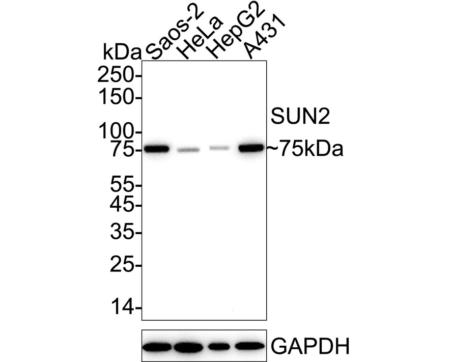
WB
Western blot analysis of SUN2 on different lysates with Rabbit anti-SUN2 antibody at 1/2,000 dilution. Lane 1: Saos-2 cell lysate, Lane 2: HeLa cell lysate, Lane 3: HepG2 cell lysate, Lane 4: A431 cell lysate, Lysates/proteins at 20 µg/Lane. Exposure time: 30 seconds; 4-20% SDS-PAGE gel. Proteins were transferred to a PVDF membrane and blocked with 5% NFDM/TBST for 1 hour at room temperature. The primary antibody at 1/2,000 dilution was used in 5% NFDM/TBST at 4℃ overnight. Goat Anti-Rabbit IgG - HRP Secondary Antibody at 1/50,000 dilution was used for 1 hour at room temperature.IHC
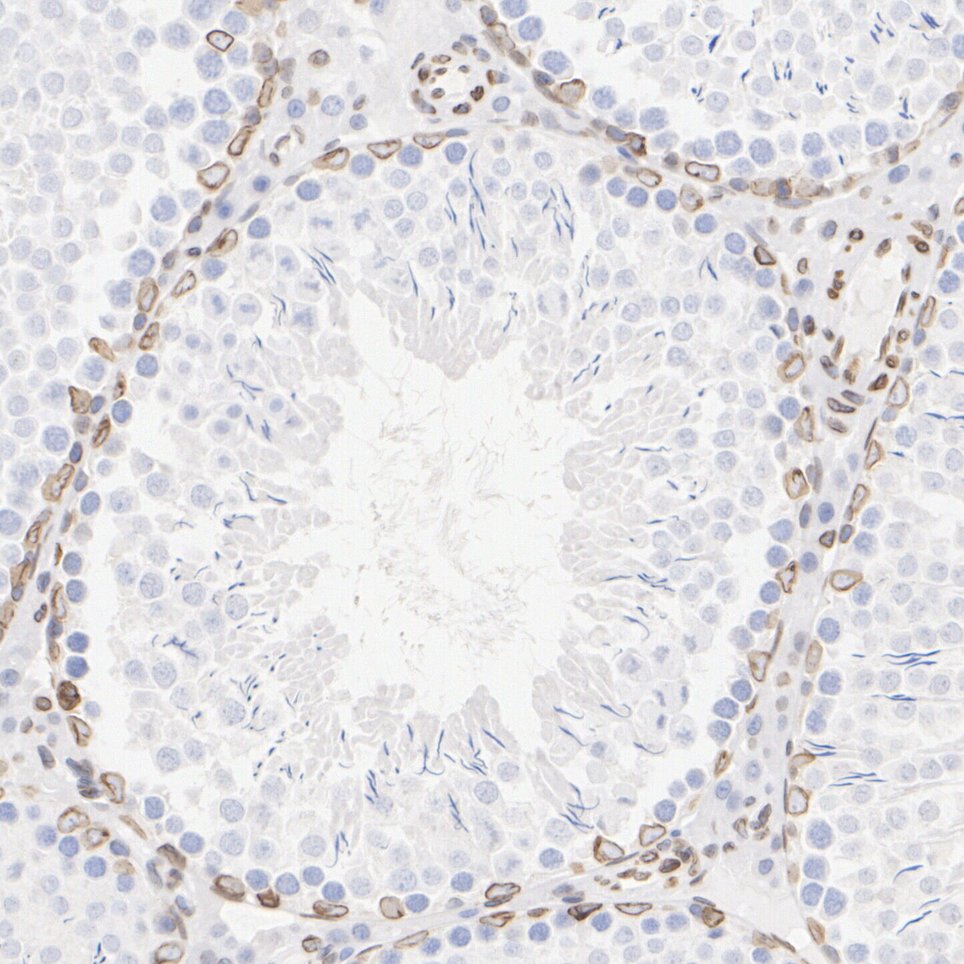
Immunohistochemical analysis of paraffin-embedded rat testis tissue with Rabbit anti-SUN2 antibody at 1/1,000 dilution. The section was pre-treated using heat mediated antigen retrieval with Tris-EDTA buffer (pH 9.0) for 20 minutes. The tissues were blocked in 1% BSA for 20 minutes at room temperature, washed with ddH2O and PBS, and then probed with the primary antibody at 1/1,000 dilution for 1 hour at room temperature. The detection was performed using an HRP conjugated compact polymer system. DAB was used as the chromogen. Tissues were counterstained with hematoxylin and mounted with DPX.ICC/IF
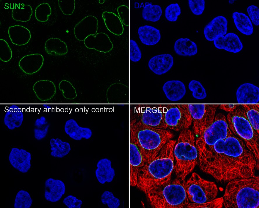
Immunocytochemistry analysis of A431 cells labeling SUN2 with Rabbit anti-SUN2 antibody at 1/50 dilution. Cells were fixed in 4% paraformaldehyde for 15 minutes at room temperature, permeabilized with 0.1% Triton X-100 in PBS for 15 minutes at room temperature, then blocked with 1% BSA in 10% negative goat serum for 1 hour at room temperature. Cells were then incubated with Rabbit anti-SUN2 antibody at 1/50 dilution in 1% BSA in PBST overnight at 4 ℃. Goat Anti-Rabbit IgG H&L (iFluor™ 488) was used as the secondary antibody at 1/1,000 dilution. PBS instead of the primary antibody was used as the secondary antibody only control. Nuclear DNA was labelled in blue with DAPI. Beta tubulin (red) was stained at 1/100 dilution overnight at +4℃. Goat Anti-Mouse IgG H&L (iFluor™ 594) was used as the secondary antibody at 1/1,000 dilution.FC
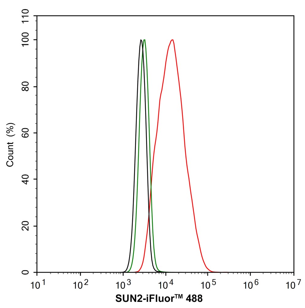
Flow cytometric analysis of A431 cells labeling SUN2. Cells were fixed and permeabilized. Then stained with the primary antibody (1/1,000) (red) compared with Rabbit IgG Isotype Control (green). After incubation of the primary antibody at +4℃ for an hour, the cells were stained with a iFluor™ 488 conjugate-Goat anti-Rabbit IgG Secondary antibody at 1/1,000 dilution for 30 minutes at +4℃. Unlabelled sample was used as a control (cells without incubation with primary antibody; black).| Product Name | SUN2 Recombinant Rabbit Monoclonal Antibody |
|---|---|
| Antibody Type | Primary Antibodies |
| Product description | SUN2 (sad1/unc-84 protein-like 2), also known as UNC84B (unc-84 homolog B), FRIGG, KIAA0668 or RAB5IP, is a 717 amino acid single-pass membrane protein that contains one SUN domain and localizes to the membrane of both the nucleus and the endosome. Widely expressed in a variety of tissues, including lung, muscle and heart, SUN2 interacts with Rab 5A and may play a role in homotypic endosome fusion. The gene encoding SUN2 maps to human chromosome 22, which houses over 500 genes and is the second smallest human chromosome. Mutations in several of the genes that map to chromosome 22 are involved in the development of Phelan-McDermid syndrome, Neurofibromatosis type 2, autism and schizophrenia. Additionally, translocations between chromosomes 9 and 22 may lead to the formation of the Philadelphia Chromosome and the subsequent production of the novel fusion protein BCR-Abl, a potent cell proliferation activator found in several types of leukemias. |
| Immunogen | Recombinant protein within human SUN2 aa 500-700. |
| Clonality | monoclonal |
|---|---|
| Isotype | IgG |
| Host Species | Recombinant rabbit |
| Tested Applications | FCICC/IFIHCWB |
| WB:1:2000 IHC:1:1000 ICC/IF:1:50-1:100 FC:1:1000 |
|
| Species Reactivity | HumanMouseRat |
| Concentration | 1mg/ml |
| Purification | Protein A |
| Alternative Names | FRIGG antibody
KIAA0668 antibody
nuclear envelope protein antibody
Protein unc-84 homolog B antibody
Rab5 interacting protein antibody
RAB5IP antibody
Sad1 and UNC84 domain containing 2 antibody
Sad1 unc-84 domain protein 2 antibody
Sad1 unc84 domain protein antibody
Sad1/unc-84 protein-like 2 antibody
Sad1/unc84 protein-like antibody
SUN domain-containing protein 2 antibody
UNC 84B antibody
unc84 homolog B (C. elegans) antibody
unc84 homolog B antibody
unc84 C. elegans homolog of B antibody UNC84B antibody |
|---|---|
| Molecular Weight(MW) | 80kDa(Observed band size: 75kDa) |
| Cellular Localization | Endosome. Nucleus inner membrane. |
WB
Western blot analysis of SUN2 on different lysates with Rabbit anti-SUN2 antibody at 1/2,000 dilution. Lane 1: Saos-2 cell lysate, Lane 2: HeLa cell lysate, Lane 3: HepG2 cell lysate, Lane 4: A431 cell lysate, Lysates/proteins at 20 µg/Lane. Exposure time: 30 seconds; 4-20% SDS-PAGE gel. Proteins were transferred to a PVDF membrane and blocked with 5% NFDM/TBST for 1 hour at room temperature. The primary antibody at 1/2,000 dilution was used in 5% NFDM/TBST at 4℃ overnight. Goat Anti-Rabbit IgG - HRP Secondary Antibody at 1/50,000 dilution was used for 1 hour at room temperature.
IHC
Immunohistochemical analysis of paraffin-embedded rat testis tissue with Rabbit anti-SUN2 antibody at 1/1,000 dilution. The section was pre-treated using heat mediated antigen retrieval with Tris-EDTA buffer (pH 9.0) for 20 minutes. The tissues were blocked in 1% BSA for 20 minutes at room temperature, washed with ddH2O and PBS, and then probed with the primary antibody at 1/1,000 dilution for 1 hour at room temperature. The detection was performed using an HRP conjugated compact polymer system. DAB was used as the chromogen. Tissues were counterstained with hematoxylin and mounted with DPX.
ICC/IF
Immunocytochemistry analysis of A431 cells labeling SUN2 with Rabbit anti-SUN2 antibody at 1/50 dilution. Cells were fixed in 4% paraformaldehyde for 15 minutes at room temperature, permeabilized with 0.1% Triton X-100 in PBS for 15 minutes at room temperature, then blocked with 1% BSA in 10% negative goat serum for 1 hour at room temperature. Cells were then incubated with Rabbit anti-SUN2 antibody at 1/50 dilution in 1% BSA in PBST overnight at 4 ℃. Goat Anti-Rabbit IgG H&L (iFluor™ 488) was used as the secondary antibody at 1/1,000 dilution. PBS instead of the primary antibody was used as the secondary antibody only control. Nuclear DNA was labelled in blue with DAPI. Beta tubulin (red) was stained at 1/100 dilution overnight at +4℃. Goat Anti-Mouse IgG H&L (iFluor™ 594) was used as the secondary antibody at 1/1,000 dilution.
FC
Flow cytometric analysis of A431 cells labeling SUN2. Cells were fixed and permeabilized. Then stained with the primary antibody (1/1,000) (red) compared with Rabbit IgG Isotype Control (green). After incubation of the primary antibody at +4℃ for an hour, the cells were stained with a iFluor™ 488 conjugate-Goat anti-Rabbit IgG Secondary antibody at 1/1,000 dilution for 30 minutes at +4℃. Unlabelled sample was used as a control (cells without incubation with primary antibody; black).| Positive Control | Rat kidney tissue lysate, rat epididymis tissue, human tonsil tissue, human colon tissue, human prostate cancer tissue, mouse liver tissue, SK-Br-3. |
|---|---|
| Application Notes | WB:1:2000 IHC:1:1000 ICC/IF:1:50-1:100 FC:1:1000 |
| Form | Liquid |
|---|---|
| Storage Instructions | Store at +4℃ after thawing. Aliquot store at -20℃. Avoid repeated freeze / thaw cycles. |
| Storage Buffer | 1*TBS (pH7.4), 1%BSA, 40%Glycerol. Preservative: 0.05% Sodium Azide. |


 Data sheet for OM643852