WB
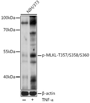
Western blot analysis of lysates from NIH/3T3 cells, using Phospho-MLKL-T357/S358/S360 pAb at 1:1000 dilution. NIH/3T3 cells were treated by TNF-α (20 ng/mL) at 37℃ for 30 minutes. Secondary antibody: HRP-conjugated Goat anti-Rabbit IgG(H+L) at 1:10000 dilution. Lysates/proteins: 25μg per lane. Blocking buffer: 3% nonfat dry milk in TBST. Detection: ECL Basic Kit. Exposure time: 60s.| Product Name | Phospho-MLKL-T357/S358/S360 Rabbit pAb |
|---|---|
| Antibody Type | Primary Antibodies |
| Immunogen | A synthetic phosphorylated peptide around T357 & S358 & S360 of human MLKL (NP_689862.1). |
| Clonality | polyclonal |
|---|---|
| Isotype | IgG |
| Host Species | Rabbit |
| Tested Applications | WB |
| WB:1:500-1:2000 |
|
| Species Reactivity | HumanMouseRat |
| Concentration | 1mg/ml |
| Purification | Affinity purified |
| Gene Symbol | MLKL |
|---|---|
| Gene Synonyms | hMLKL |
| Gene Full Name | mixed lineage kinase domain like pseudokinase |
| Gene Summary | This gene belongs to the protein kinase superfamily. The encoded protein contains a protein kinase-like domain; however, is thought to be inactive because it lacks several residues required for activity. This protein plays a critical role in tumor necrosis factor (TNF)-induced necroptosis, a programmed cell death process, via interaction with receptor-interacting protein 3 (RIP3), which is a key signaling molecule in necroptosis pathway. Inhibitor studies and knockdown of this gene inhibited TNF-induced necrosis. High levels of this protein and RIP3 are associated with inflammatory bowel disease in children. Alternatively spliced transcript variants have been described for this gene. [provided by RefSeq, Sep 2015] |
| Molecular Weight(MW) | 54kDa |
| Cellular Localization | Cell membrane, Cytoplasm. |
WB
Western blot analysis of lysates from NIH/3T3 cells, using Phospho-MLKL-T357/S358/S360 pAb at 1:1000 dilution. NIH/3T3 cells were treated by TNF-α (20 ng/mL) at 37℃ for 30 minutes. Secondary antibody: HRP-conjugated Goat anti-Rabbit IgG(H+L) at 1:10000 dilution. Lysates/proteins: 25μg per lane. Blocking buffer: 3% nonfat dry milk in TBST. Detection: ECL Basic Kit. Exposure time: 60s.| Application Notes | WB:1:500-1:2000 |
|---|
| Form | Liquid |
|---|---|
| Storage Instructions | Store at -20℃. Avoid freeze / thaw cycles. |
| Storage Buffer | Buffer: PBS with 0.05% proclin300, 50% glycerol, pH7.3. |


 Data sheet for OM643854