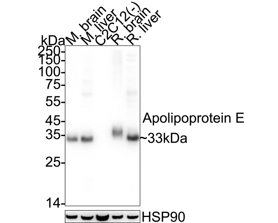

WB
Western blot analysis of Apolipoprotein E on different lysates with Rabbit anti-Apolipoprotein E antibody at 1/5,000 dilution. Lane 1: Mouse brain tissue lysate, Lane 2: Mouse liver tissue lysate, Lane 3: C2C12 cell lysate (negative), Lane 4: Rat brain tissue lysate, Lane 5: Rat liver tissue lysate, Lysates/proteins at 20 µg/Lane. Exposure time: 10 seconds; 4-20% SDS-PAGE gel. Proteins were transferred to a PVDF membrane and blocked with 5% NFDM/TBST for 1 hour at room temperature. The primary antibody at 1/5,000 dilution was used in primary antibody dilution at 4℃ overnight. Goat Anti-Rabbit IgG - HRP Secondary Antibody at 1/50,000 dilution was used for 1 hour at room temperature.IHC
Immunohistochemical analysis of paraffin-embedded mouse brain tissue with Rabbit anti-Apolipoprotein E antibody at 1/2,000 dilution. The section was pre-treated using heat mediated antigen retrieval with Tris-EDTA buffer (pH 9.0) for 20 minutes. The tissues were blocked in 1% BSA for 20 minutes at room temperature, washed with ddH2O and PBS, and then probed with the primary antibody at 1/2,000 dilution for 1 hour at room temperature. The detection was performed using an HRP conjugated compact polymer system. DAB was used as the chromogen. Tissues were counterstained with hematoxylin and mounted with DPX.IF-F
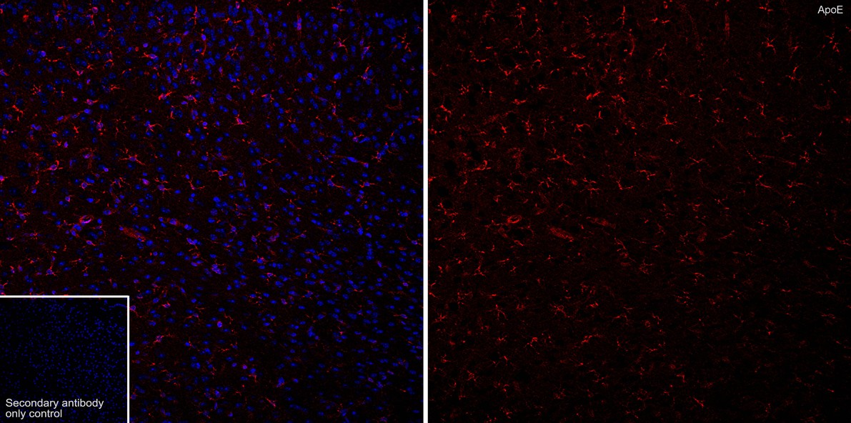
Immunofluorescence analysis of frozen mouse brain tissue with Rabbit anti-Apolipoprotein E antibody at 1/500 dilution. The section was not undergone antigen retrieval. The tissues were blocked in 10% negative goat serum for 1 hour at room temperature, washed with PBS, and then probed with the primary antibody (red) at 1/500 dilution overnight at 4 ℃, washed with PBS. Goat Anti-Rabbit IgG H&L (iFluor™ 594) was used as the secondary antibody at 1/1,000 dilution. Nuclei were counterstained with DAPI (blue).| Product Name | Apolipoprotein E Recombinant Rabbit Monoclonal Antibody |
|---|---|
| Antibody Type | Primary Antibodies |
| Clonality | monoclonal |
|---|---|
| Isotype | IgG |
| Host Species | Rabbit |
| Tested Applications | IF-FIHCWB |
| WB:1:5000 IHC:1:2000 IF-F:1:500 |
|
| Species Reactivity | MouseRat |
| Concentration | 1mg/ml |
| Purification | Protein A |
| Gene Symbol | Apoe |
|---|---|
| Gene Synonyms | AD2, APO-E, ApoE4, LDLCQ5, LPG |
| Gene Full Name | apolipoprotein E |
| Gene Summary | This gene encodes a member of the apolipoprotein A1/A4/E family of proteins. This protein is involved in the transport of lipoproteins in the blood. It binds to a specific liver and peripheral cell receptor, and is essential for the normal catabolism of triglyceride-rich lipoprotein constituents. Homozygous knockout mice for this gene accumulate high levels of cholesterol in the blood and develop atherosclerosis. Different alleles of this gene have been associated with either increased risk or a protective effect for Alzheimer's disease in human patients. This gene maps to chromosome 7 in a cluster with the related apolipoprotein C1, C2 and C4 genes. [provided by RefSeq, Apr 2015] |
| Molecular Weight(MW) | 36kDa(Observed band size: 33kDa) |
| Cellular Localization | Secreted, extracellular space, extracellular matrix. |
WB
Western blot analysis of Apolipoprotein E on different lysates with Rabbit anti-Apolipoprotein E antibody at 1/5,000 dilution. Lane 1: Mouse brain tissue lysate, Lane 2: Mouse liver tissue lysate, Lane 3: C2C12 cell lysate (negative), Lane 4: Rat brain tissue lysate, Lane 5: Rat liver tissue lysate, Lysates/proteins at 20 µg/Lane. Exposure time: 10 seconds; 4-20% SDS-PAGE gel. Proteins were transferred to a PVDF membrane and blocked with 5% NFDM/TBST for 1 hour at room temperature. The primary antibody at 1/5,000 dilution was used in primary antibody dilution at 4℃ overnight. Goat Anti-Rabbit IgG - HRP Secondary Antibody at 1/50,000 dilution was used for 1 hour at room temperature.
IHC
Immunohistochemical analysis of paraffin-embedded mouse brain tissue with Rabbit anti-Apolipoprotein E antibody at 1/2,000 dilution. The section was pre-treated using heat mediated antigen retrieval with Tris-EDTA buffer (pH 9.0) for 20 minutes. The tissues were blocked in 1% BSA for 20 minutes at room temperature, washed with ddH2O and PBS, and then probed with the primary antibody at 1/2,000 dilution for 1 hour at room temperature. The detection was performed using an HRP conjugated compact polymer system. DAB was used as the chromogen. Tissues were counterstained with hematoxylin and mounted with DPX.
IF-F
Immunofluorescence analysis of frozen mouse brain tissue with Rabbit anti-Apolipoprotein E antibody at 1/500 dilution. The section was not undergone antigen retrieval. The tissues were blocked in 10% negative goat serum for 1 hour at room temperature, washed with PBS, and then probed with the primary antibody (red) at 1/500 dilution overnight at 4 ℃, washed with PBS. Goat Anti-Rabbit IgG H&L (iFluor™ 594) was used as the secondary antibody at 1/1,000 dilution. Nuclei were counterstained with DAPI (blue).| Application Notes | WB:1:5000 IHC:1:2000 IF-F:1:500 |
|---|
| Form | Liquid |
|---|---|
| Storage Instructions | Store at +4℃ after thawing. Aliquot store at -20℃ or -80℃. Avoid repeated freeze / thaw cycles. |
| Storage Buffer | 1*TBS (pH7.4), 0.05% BSA, 40% Glycerol. Preservative: 0.05% Sodium Azide. |


 Data sheet for OM643901