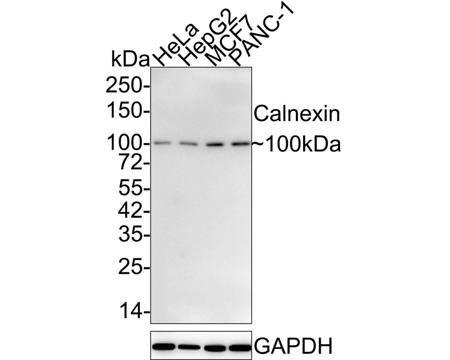
WB
Western blot analysis of Calnexin on different lysates with Rabbit anti-Calnexin antibody at 1/2,000 dilution. Lane 1: HeLa cell lysate, Lane 2: HepG2 cell lysate, Lane 3: MCF7 cell lysate, Lane 4: PANC-1 cell lysate, Lysates/proteins at 15 µg/Lane. Exposure time: 2 minutes; 4-20% SDS-PAGE gel. Proteins were transferred to a PVDF membrane and blocked with 5% NFDM/TBST for 1 hour at room temperature. The primary antibody at 1/2,000 dilution was used in 5% NFDM/TBST at 4℃ overnight. Goat Anti-Rabbit IgG - HRP Secondary Antibody at 1/50,000 dilution was used for 1 hour at room temperature.IHC
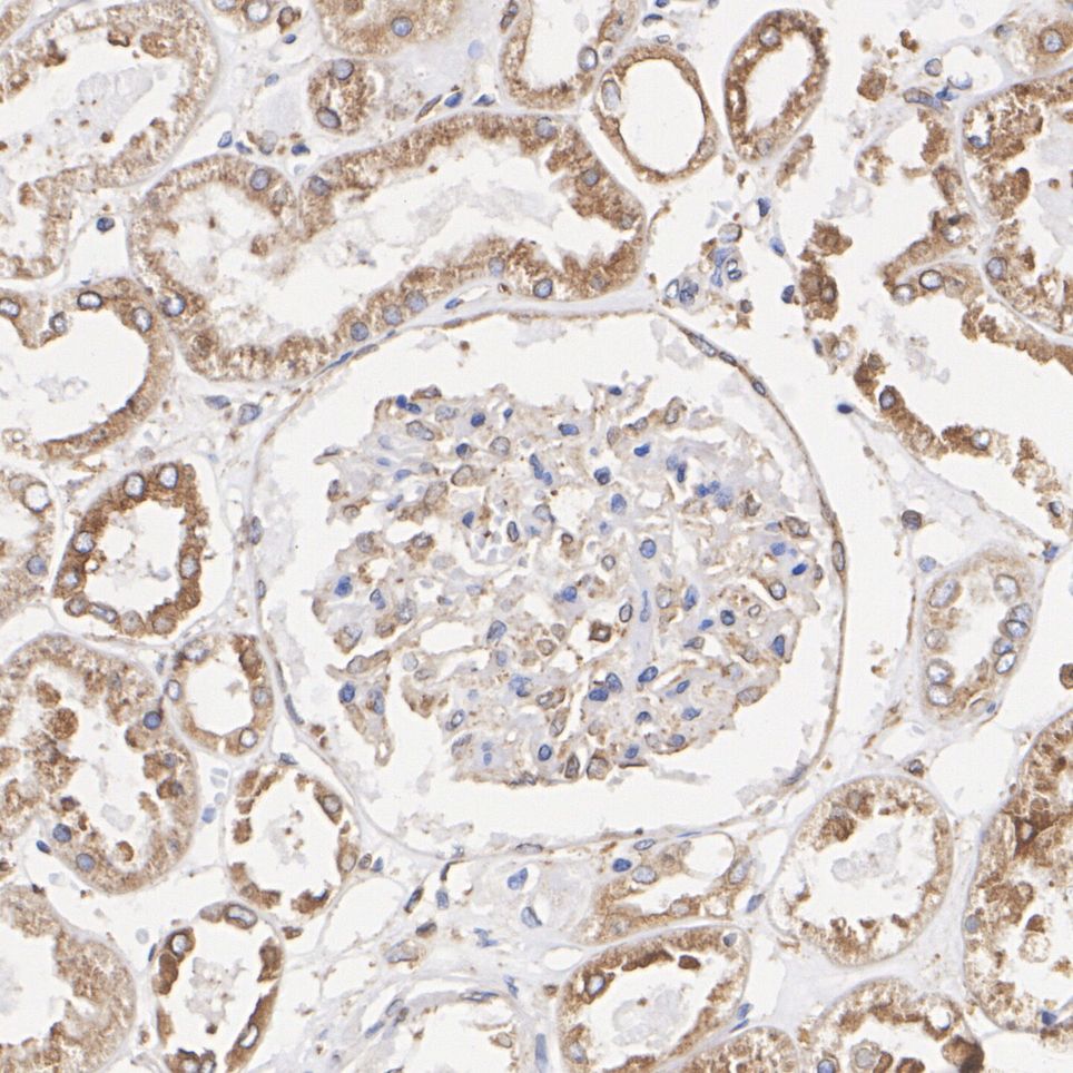
Immunohistochemical analysis of paraffin-embedded human kidney tissue with Rabbit anti-Calnexin antibody at 1/5,000 dilution. The section was pre-treated using heat mediated antigen retrieval with Tris-EDTA buffer (pH 9.0) for 20 minutes. The tissues were blocked in 1% BSA for 20 minutes at room temperature, washed with ddH2O and PBS, and then probed with the primary antibody at 1/5,000 dilution for 1 hour at room temperature. The detection was performed using an HRP conjugated compact polymer system. DAB was used as the chromogen. Tissues were counterstained with hematoxylin and mounted with DPX.IP
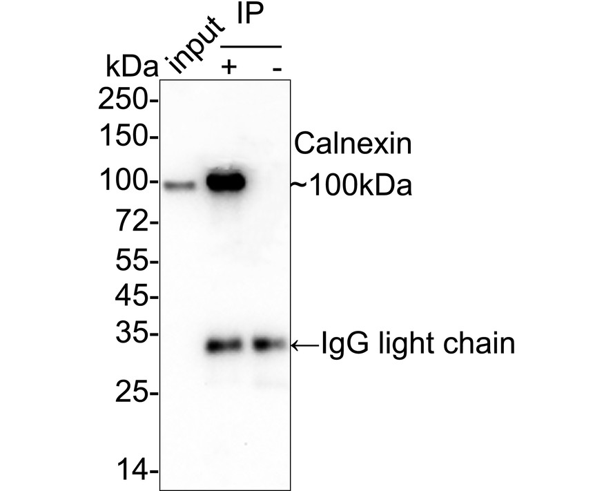
Calnexin was immunoprecipitated from 0.2 mg HeLa cell lysate with Rabbit anti-Calnexin antibody at 2µg/25µl agarose. Western blot was performed from the immunoprecipitate using Rabbit anti-Calnexin antibody at 1/1,000 dilution. Anti-Rabbit IgG for IP Nano-secondary antibody at 1/5,000 dilution was used for 1 hour at room temperature. Lane 1: HeLa cell lysate (input), Lane 2: Rabbit anti-Calnexin antibody IP in HeLa cell lysate, Lane 3: Rabbit IgG instead of Rabbit anti-Calnexin antibody in HeLa cell lysate, Blocking/Dilution buffer: 5% NFDM/TBST. Exposure time: 1 minute 5 seconds.| Product Name | Calnexin Recombinant Rabbit Monoclonal Antibody |
|---|---|
| Antibody Type | Primary Antibodies |
| Immunogen | Synthetic peptide within Human Calnexin aa 543-592 / 592. |
| Clonality | monoclonal |
|---|---|
| Isotype | IgG |
| Host Species | Rabbit |
| Tested Applications | IHCIPWB |
| WB:1:1000-1:2000 IHC:1:1000-1:5000 IP:1-2μg/sample |
|
| Species Reactivity | HumanMouseRat |
| Concentration | 1mg/ml |
| Purification | Protein A |
| Gene Symbol | CANX |
|---|---|
| Gene Synonyms | CNX P90 IP90 |
| Gene Full Name | calnexin |
| Gene Summary | This gene encodes a member of the calnexin family of molecular chaperones. The encoded protein is a calcium-binding, endoplasmic reticulum (ER)-associated protein that interacts transiently with newly synthesized N-linked glycoproteins, facilitating protein folding and assembly. It may also play a central role in the quality control of protein folding by retaining incorrectly folded protein subunits within the ER for degradation. Alternatively spliced transcript variants encoding different isoforms have been described. [provided by RefSeq, Jun 2018] |
| Molecular Weight(MW) | 68kDa(Observed band size: 100kDa) |
| Cellular Localization | Endoplasmic reticulum membrane, Endoplasmic reticulum, Melanosome. |
WB
Western blot analysis of Calnexin on different lysates with Rabbit anti-Calnexin antibody at 1/2,000 dilution. Lane 1: HeLa cell lysate, Lane 2: HepG2 cell lysate, Lane 3: MCF7 cell lysate, Lane 4: PANC-1 cell lysate, Lysates/proteins at 15 µg/Lane. Exposure time: 2 minutes; 4-20% SDS-PAGE gel. Proteins were transferred to a PVDF membrane and blocked with 5% NFDM/TBST for 1 hour at room temperature. The primary antibody at 1/2,000 dilution was used in 5% NFDM/TBST at 4℃ overnight. Goat Anti-Rabbit IgG - HRP Secondary Antibody at 1/50,000 dilution was used for 1 hour at room temperature.
IHC
Immunohistochemical analysis of paraffin-embedded human kidney tissue with Rabbit anti-Calnexin antibody at 1/5,000 dilution. The section was pre-treated using heat mediated antigen retrieval with Tris-EDTA buffer (pH 9.0) for 20 minutes. The tissues were blocked in 1% BSA for 20 minutes at room temperature, washed with ddH2O and PBS, and then probed with the primary antibody at 1/5,000 dilution for 1 hour at room temperature. The detection was performed using an HRP conjugated compact polymer system. DAB was used as the chromogen. Tissues were counterstained with hematoxylin and mounted with DPX.
IP
Calnexin was immunoprecipitated from 0.2 mg HeLa cell lysate with Rabbit anti-Calnexin antibody at 2µg/25µl agarose. Western blot was performed from the immunoprecipitate using Rabbit anti-Calnexin antibody at 1/1,000 dilution. Anti-Rabbit IgG for IP Nano-secondary antibody at 1/5,000 dilution was used for 1 hour at room temperature. Lane 1: HeLa cell lysate (input), Lane 2: Rabbit anti-Calnexin antibody IP in HeLa cell lysate, Lane 3: Rabbit IgG instead of Rabbit anti-Calnexin antibody in HeLa cell lysate, Blocking/Dilution buffer: 5% NFDM/TBST. Exposure time: 1 minute 5 seconds.| Application Notes | WB:1:1000-1:2000 IHC:1:1000-1:5000 IP:1-2μg/sample |
|---|
| Form | Liquid |
|---|---|
| Storage Instructions | Store at +4℃ after thawing. Aliquot store at -20℃ or -80℃. Avoid repeated freeze / thaw cycles. |
| Storage Buffer | 1*TBS (pH7.4), 0.05% BSA, 40% Glycerol. Preservative: 0.05% Sodium Azide. |


 Data sheet for OM643906