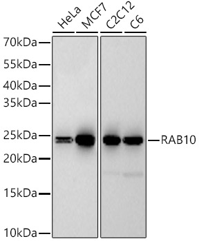
WB
Western blot analysis of various lysates, using RAB10 Rabbit mAb at 1:60000 dilution. Secondary antibody: HRP Goat Anti-Rabbit IgG (H+L) at 1:10000 dilution. Lysates/proteins: 25μg per lane. Blocking buffer: 3% nonfat dry milk in TBST. Detection: ECL Basic Kit. Exposure time: 180sICC/IF
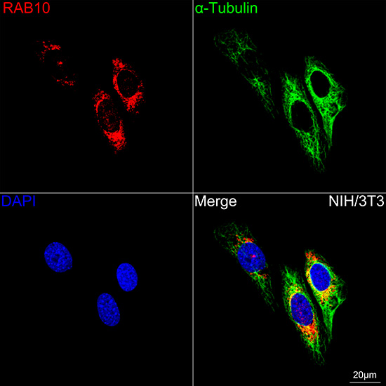
Confocal imaging of NIH/3T3 cells using RAB10 Rabbit mAb (dilution 1:200) followed by a further incubation with Cy3 Goat Anti-Rabbit IgG (H+L) (dilution 1:500) (Red). The cells were counterstained with α-Tubulin Mouse mAb (dilution 1:400) followed by incubation with 488-conjugated Goat Anti-Mouse IgG (H+L) Ab (dilution 1:500) (Green). DAPI was used for nuclear staining (Blue). Objective: 100x.| Product Name | RAB10 Rabbit mAb |
|---|---|
| Antibody Type | Primary Antibodies |
| Immunogen | A synthetic peptide corresponding to a sequence within amino acids 101-200 of human RAB10 (NP_057215.3). |
| Clonality | monoclonal |
|---|---|
| Isotype | IgG |
| Host Species | Rabbit |
| Tested Applications | ICC/IFWB |
| WB:1:10000-1:60000 ICC/IF:1:50-1:200 |
|
| Species Reactivity | HumanMouseRat |
| Concentration | 1mg/ml |
| Purification | Affinity purified |
| Gene Symbol | RAB10 |
|---|---|
| Gene Full Name | RAB10, member RAS oncogene family |
| Gene Summary | RAB10 belongs to the RAS (see HRAS; MIM 190020) superfamily of small GTPases. RAB proteins localize to exocytic and endocytic compartments and regulate intracellular vesicle trafficking (Bao et al., 1998 [PubMed 9918381]).[supplied by OMIM, Mar 2009] |
| Molecular Weight(MW) | 23kDa |
| Cellular Localization | Cell projection, Cytoplasmic side, Cytoplasmic vesicle, Cytoplasmic vesicle membrane, Endoplasmic reticulum membrane, Endosome membrane, Golgi apparatus, Lipid-anchor, Recycling endosome membrane, cilium, phagosome membrane, trans-Golgi network membrane. |
WB
Western blot analysis of various lysates, using RAB10 Rabbit mAb at 1:60000 dilution. Secondary antibody: HRP Goat Anti-Rabbit IgG (H+L) at 1:10000 dilution. Lysates/proteins: 25μg per lane. Blocking buffer: 3% nonfat dry milk in TBST. Detection: ECL Basic Kit. Exposure time: 180s
ICC/IF
Confocal imaging of NIH/3T3 cells using RAB10 Rabbit mAb (dilution 1:200) followed by a further incubation with Cy3 Goat Anti-Rabbit IgG (H+L) (dilution 1:500) (Red). The cells were counterstained with α-Tubulin Mouse mAb (dilution 1:400) followed by incubation with 488-conjugated Goat Anti-Mouse IgG (H+L) Ab (dilution 1:500) (Green). DAPI was used for nuclear staining (Blue). Objective: 100x.| Application Notes | WB:1:10000-1:60000 ICC/IF:1:50-1:200 |
|---|
| Form | Liquid |
|---|---|
| Storage Instructions | Store at -20℃. Avoid freeze / thaw cycles. |
| Storage Buffer | Buffer: PBS with 0.05% proclin300, 0.05% BSA, 50% glycerol, pH7.3. |


 Data sheet for OM643957