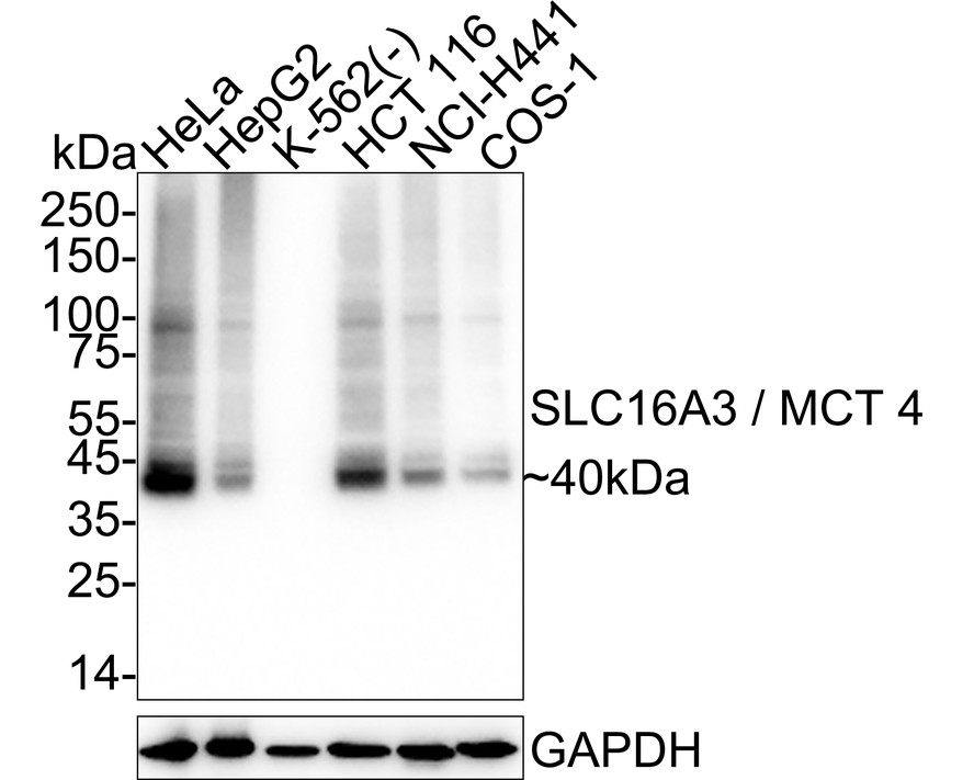
WB
Western blot analysis of SLC16A3 / MCT 4 on different lysates with Rabbit anti-SLC16A3 / MCT 4 antibody at 1/20,000 dilution. Lane 1: HeLa cell lysate, Lane 2: HepG2 cell lysate, Lane 3: K-562 cell lysate (negative), Lane 4: HCT 116 cell lysate, Lane 5: NCI-H441 cell lysate, Lane 6: COS-1 cell lysate, Lysates/proteins at 20 µg/Lane. Exposure time: 6 seconds; 4-20% SDS-PAGE gel. Proteins were transferred to a PVDF membrane and blocked with 5% NFDM/TBST for 1 hour at room temperature. The primary antibody at 1/20,000 dilution was used in 5% NFDM/TBST at 4℃ overnight. Goat Anti-Rabbit IgG - HRP Secondary Antibody at 1/50,000 dilution was used for 1 hour at room temperature.IHC
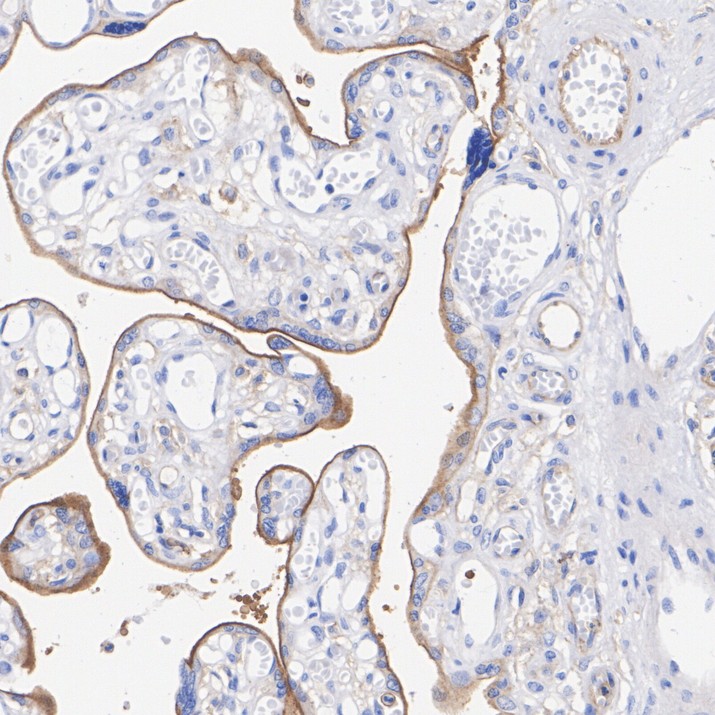
Immunohistochemical analysis of paraffin-embedded human placenta tissue with Rabbit anti-SLC16A3 / MCT 4 antibody at 1/1,000 dilution. The section was pre-treated using heat mediated antigen retrieval with Tris-EDTA buffer (pH 9.0) for 20 minutes. The tissues were blocked in 1% BSA for 20 minutes at room temperature, washed with ddH2O and PBS, and then probed with the primary antibody at 1/1,000 dilution for 1 hour at room temperature. The detection was performed using an HRP conjugated compact polymer system. DAB was used as the chromogen. Tissues were counterstained with hematoxylin and mounted with DPX.ICC/IF
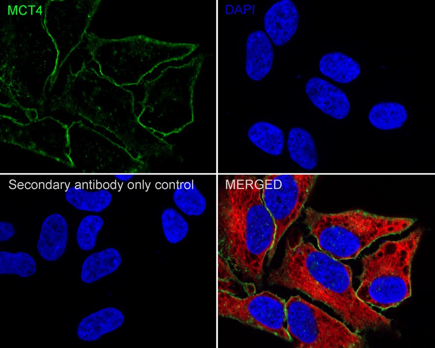
Immunocytochemistry analysis of HeLa cells labeling SLC16A3 / MCT 4 with Rabbit anti-SLC16A3 / MCT 4 antibody at 1/100 dilution. Cells were fixed in 4% paraformaldehyde for 20 minutes at room temperature, permeabilized with 0.1% Triton X-100 in PBS for 5 minutes at room temperature, then blocked with 1% BSA in 10% negative goat serum for 1 hour at room temperature. Cells were then incubated with Rabbit anti-SLC16A3 / MCT 4 antibody at 1/100 dilution in 1% BSA in PBST overnight at 4 ℃. Goat Anti-Rabbit IgG H&L (iFluor™ 488) was used as the secondary antibody at 1/1,000 dilution. PBS instead of the primary antibody was used as the secondary antibody only control. Nuclear DNA was labelled in blue with DAPI. Beta tubulin (red) was stained at 1/100 dilution overnight at +4℃. Goat Anti-Mouse IgG H&L (iFluor™ 594) was used as the secondary antibody at 1/1,000 dilution.FC
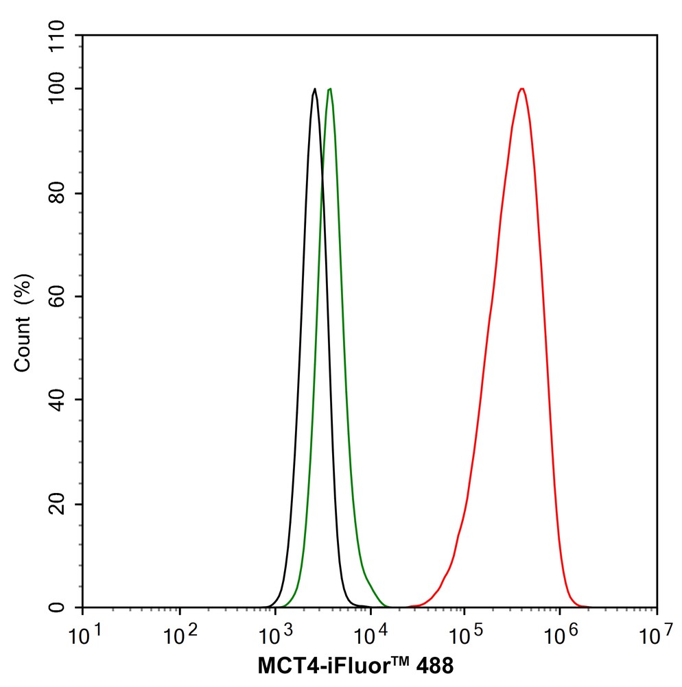
Flow cytometric analysis of HeLa cells labeling SLC16A3 / MCT 4. Cells were fixed and permeabilized. Then stained with the primary antibody (1/1,000) (red) compared with Rabbit IgG Isotype Control (green). After incubation of the primary antibody at +4℃ for an hour, the cells were stained with a iFluor™ 488 conjugate-Goat anti-Rabbit IgG Secondary antibody at 1/1,000 dilution for 30 minutes at +4℃. Unlabelled sample was used as a control (cells without incubation with primary antibody; black).| Product Name | SLC16A3 / MCT 4 Recombinant Rabbit Monoclonal Antibody |
|---|---|
| Antibody Type | Primary Antibodies |
| Clonality | monoclonal |
|---|---|
| Isotype | IgG |
| Host Species | Rabbit |
| Tested Applications | FCICC/IFIHCWB |
| WB:1:20000 IHC:1:1000 ICC/IF:1:100 FC:1:1000 |
|
| Species Reactivity | HumanMonkey |
| Concentration | 1mg/ml |
| Purification | Protein A |
| Gene Symbol | SLC16A3 |
|---|---|
| Gene Synonyms | MCT3 MCT4 MCT 3 MCT 4 MCT-3 MCT-4 |
| Gene Full Name | solute carrier family 16 member 3 |
| Gene Summary | Lactic acid and pyruvate transport across plasma membranes is catalyzed by members of the proton-linked monocarboxylate transporter (MCT) family, which has been designated solute carrier family-16. Each MCT appears to have slightly different substrate and inhibitor specificities and transport kinetics, which are related to the metabolic requirements of the tissues in which it is found. The MCTs, which include MCT1 (SLC16A1; MIM 600682) and MCT2 (SLC16A7; MIM 603654), are characterized by 12 predicted transmembrane domains (Price et al., 1998 [PubMed 9425115]).[supplied by OMIM, Mar 2008] |
| Molecular Weight(MW) | 49kDa(Observed band size: 40 kDa) |
| Cellular Localization | Cell membrane, Basolateral cell membrane. |
WB
Western blot analysis of SLC16A3 / MCT 4 on different lysates with Rabbit anti-SLC16A3 / MCT 4 antibody at 1/20,000 dilution. Lane 1: HeLa cell lysate, Lane 2: HepG2 cell lysate, Lane 3: K-562 cell lysate (negative), Lane 4: HCT 116 cell lysate, Lane 5: NCI-H441 cell lysate, Lane 6: COS-1 cell lysate, Lysates/proteins at 20 µg/Lane. Exposure time: 6 seconds; 4-20% SDS-PAGE gel. Proteins were transferred to a PVDF membrane and blocked with 5% NFDM/TBST for 1 hour at room temperature. The primary antibody at 1/20,000 dilution was used in 5% NFDM/TBST at 4℃ overnight. Goat Anti-Rabbit IgG - HRP Secondary Antibody at 1/50,000 dilution was used for 1 hour at room temperature.
IHC
Immunohistochemical analysis of paraffin-embedded human placenta tissue with Rabbit anti-SLC16A3 / MCT 4 antibody at 1/1,000 dilution. The section was pre-treated using heat mediated antigen retrieval with Tris-EDTA buffer (pH 9.0) for 20 minutes. The tissues were blocked in 1% BSA for 20 minutes at room temperature, washed with ddH2O and PBS, and then probed with the primary antibody at 1/1,000 dilution for 1 hour at room temperature. The detection was performed using an HRP conjugated compact polymer system. DAB was used as the chromogen. Tissues were counterstained with hematoxylin and mounted with DPX.
ICC/IF
Immunocytochemistry analysis of HeLa cells labeling SLC16A3 / MCT 4 with Rabbit anti-SLC16A3 / MCT 4 antibody at 1/100 dilution. Cells were fixed in 4% paraformaldehyde for 20 minutes at room temperature, permeabilized with 0.1% Triton X-100 in PBS for 5 minutes at room temperature, then blocked with 1% BSA in 10% negative goat serum for 1 hour at room temperature. Cells were then incubated with Rabbit anti-SLC16A3 / MCT 4 antibody at 1/100 dilution in 1% BSA in PBST overnight at 4 ℃. Goat Anti-Rabbit IgG H&L (iFluor™ 488) was used as the secondary antibody at 1/1,000 dilution. PBS instead of the primary antibody was used as the secondary antibody only control. Nuclear DNA was labelled in blue with DAPI. Beta tubulin (red) was stained at 1/100 dilution overnight at +4℃. Goat Anti-Mouse IgG H&L (iFluor™ 594) was used as the secondary antibody at 1/1,000 dilution.
FC
Flow cytometric analysis of HeLa cells labeling SLC16A3 / MCT 4. Cells were fixed and permeabilized. Then stained with the primary antibody (1/1,000) (red) compared with Rabbit IgG Isotype Control (green). After incubation of the primary antibody at +4℃ for an hour, the cells were stained with a iFluor™ 488 conjugate-Goat anti-Rabbit IgG Secondary antibody at 1/1,000 dilution for 30 minutes at +4℃. Unlabelled sample was used as a control (cells without incubation with primary antibody; black).| Application Notes | WB:1:20000 IHC:1:1000 ICC/IF:1:100 FC:1:1000 |
|---|
| Form | Liquid |
|---|---|
| Storage Instructions | Store at +4℃ after thawing. Aliquot store at -20℃. Avoid repeated freeze / thaw cycles. |
| Storage Buffer | PBS (pH7.4), 0.1% BSA, 40% Glycerol. Preservative: 0.05% Sodium Azide. |


 Data sheet for OM643966