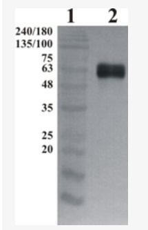
WB
Western-Blot detection of human GFRa-1, using Chicken polyclonal antibody to human GFRa-1. GFRa-1 was C-terminally His tagged. Protein was expressed by CHO cell culture. 100 ng of purified protein was loaded per lane. Lane 1. Prestained protein ladder. Liae 2. hGFRa-1. Primary antibody dilution 1:5000 was used. Rabbit anti-Chicken antibody was used as secondary antibody.ICC/IF
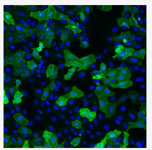
Immunofluorescence detection of human GFRa-1 expressed in U2OS cells. GFRa-1 was visualized using Chicken polyclonal antibody to human GFRa-1 (dilution 1:2000) and Goat anti-chicken IgY - H&L DyLight 550 (dilution 1:200) as secondary antibody. For nuclear staining DAPI was used. ArrayScan VTI platform (Thermo Scientific) was used for image acquisition (10x objective). Composite picture was generated using pseudocolors green for GFRa-1 specific signal and blue for nuclei.| Product Name | GDNF Receptor alpha 1 (GFRA1) Chicken Polyclonal Antibody |
|---|---|
| Antibody Type | Primary Antibodies |
| Immunogen | Recombinant His-tagged human GFRa-1 protein produced using CHO-based. For production of hGFRa-1, glycosylphosphatidyl-inositol GPI-anchor was removed and protein was secreted to the cell culture supernatant. Protein was purified by Ni-affinity chromatography following gel-filtration from cell culture supernatant. |
| Clonality | polyclonal |
|---|---|
| Isotype | IgY |
| Host Species | Chicken |
| Tested Applications | ICC/IFWB |
| WB:1:5000-1:10000 ICC/IF:1:1000-1:2000 |
|
| Species Reactivity | Human |
| Concentration | 1mg/ml |
| Purification | Affinity purified |
| Gene Symbol | GFRA1 |
|---|---|
| Gene Synonyms | GDNFR RET1L RETL1 RHDA4 TRNR1 GDNFRA GFRalpha-1 GFR-ALPHA-1 GDNFR-alpha-1 |
| Gene Full Name | GDNF family receptor alpha 1 |
| Gene Summary | This gene encodes a member of the glial cell line-derived neurotrophic factor receptor (GDNFR) family of proteins. The encoded preproprotein is proteolytically processed to generate the mature receptor. Glial cell line-derived neurotrophic factor (GDNF) and neurturin (NTN) are two structurally related, potent neurotrophic factors that play key roles in the control of neuron survival and differentiation. This receptor is a glycosylphosphatidylinositol (GPI)-linked cell surface receptor for both GDNF and NTN, and mediates activation of the RET tyrosine kinase receptor. This gene is a candidate gene for Hirschsprung disease. Alternative splicing results in multiple transcript variants, at least one of which encodes a preproprotein that is proteolytically processed. [provided by RefSeq, Jan 2016] |
| Molecular Weight(MW) | 51kDa |
| Cellular Localization | Cell membrane,Endosome,Golgi apparatus,Membrane. |
WB
Western-Blot detection of human GFRa-1, using Chicken polyclonal antibody to human GFRa-1. GFRa-1 was C-terminally His tagged. Protein was expressed by CHO cell culture. 100 ng of purified protein was loaded per lane. Lane 1. Prestained protein ladder. Liae 2. hGFRa-1. Primary antibody dilution 1:5000 was used. Rabbit anti-Chicken antibody was used as secondary antibody.
ICC/IF
Immunofluorescence detection of human GFRa-1 expressed in U2OS cells. GFRa-1 was visualized using Chicken polyclonal antibody to human GFRa-1 (dilution 1:2000) and Goat anti-chicken IgY - H&L DyLight 550 (dilution 1:200) as secondary antibody. For nuclear staining DAPI was used. ArrayScan VTI platform (Thermo Scientific) was used for image acquisition (10x objective). Composite picture was generated using pseudocolors green for GFRa-1 specific signal and blue for nuclei.| Application Notes | WB:1:5000-1:10000 ICC/IF:1:1000-1:2000 |
|---|
| Form | Liquid |
|---|---|
| Storage Instructions | Upon receipt, store at 2-8°C. As product is (NH4)2SO4 precipitate, mix well by pipetting or vortexing prior use. |
| Storage Buffer | Concentrated Ammonium Sulphate in PBS pH 7.4 State: Aff - Purified State: Liquid purified IgY fraction |


 Data sheet for OM643974