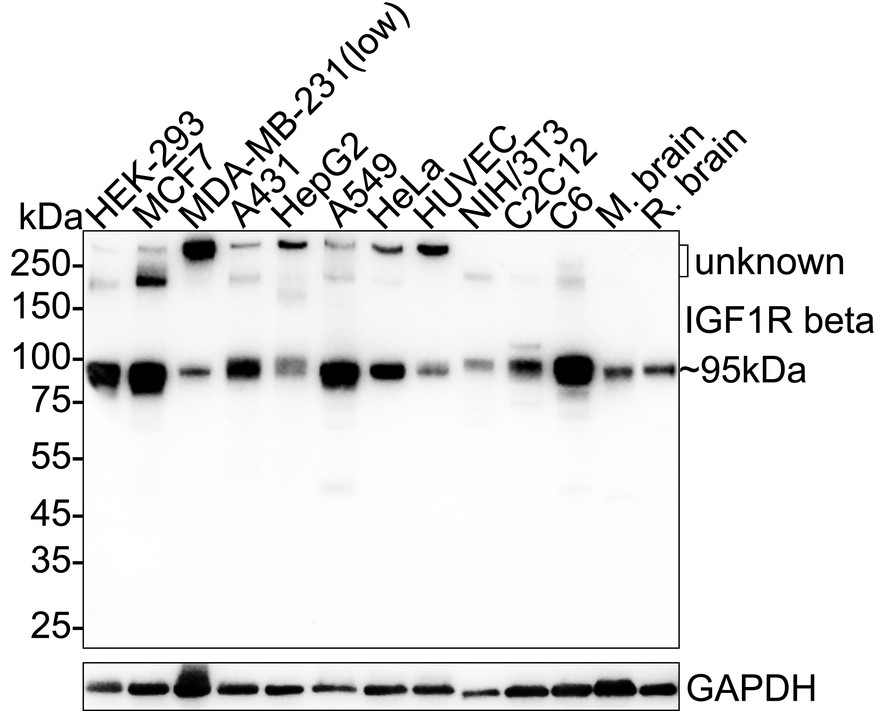
WB
Western blot analysis of IGF1 Receptor beta on different lysates with Rabbit anti-IGF1 Receptor beta antibody at 1/2,000 dilution. Lane 1: HEK-293 cell lysate (20 µg/Lane), Lane 2: MCF7 cell lysate (20 µg/Lane), Lane 3: MDA-MB-231 cell lysate (low expression) (20 µg/Lane), Lane 4: A431 cell lysate (20 µg/Lane), Lane 5: HepG2 cell lysate (20 µg/Lane), Lane 6: A549 cell lysate (20 µg/Lane), Lane 7: HeLa cell lysate (20 µg/Lane), Lane 8: HUVEC cell lysate (20 µg/Lane), Lane 9: NIH/3T3 cell lysate (20 µg/Lane), Lane 10: C2C12 cell lysate (20 µg/Lane), Lane 11: C6 cell lysate (20 µg/Lane), Lane 12: Mouse brain tissue lysate (40 µg/Lane), Lane 13: Rat brain tissue lysate (40 µg/Lane), Exposure time: 3 minutes; 4-20% SDS-PAGE gel. Proteins were transferred to a PVDF membrane and blocked with 5% NFDM/TBST for 1 hour at room temperature. The primary antibody at 1/2,000 dilution was used in 5% NFDM/TBST at 4℃ overnight. Goat Anti-Rabbit IgG - HRP Secondary Antibody at 1/50,000 dilution was used for 1 hour at room temperature.ICC/IF
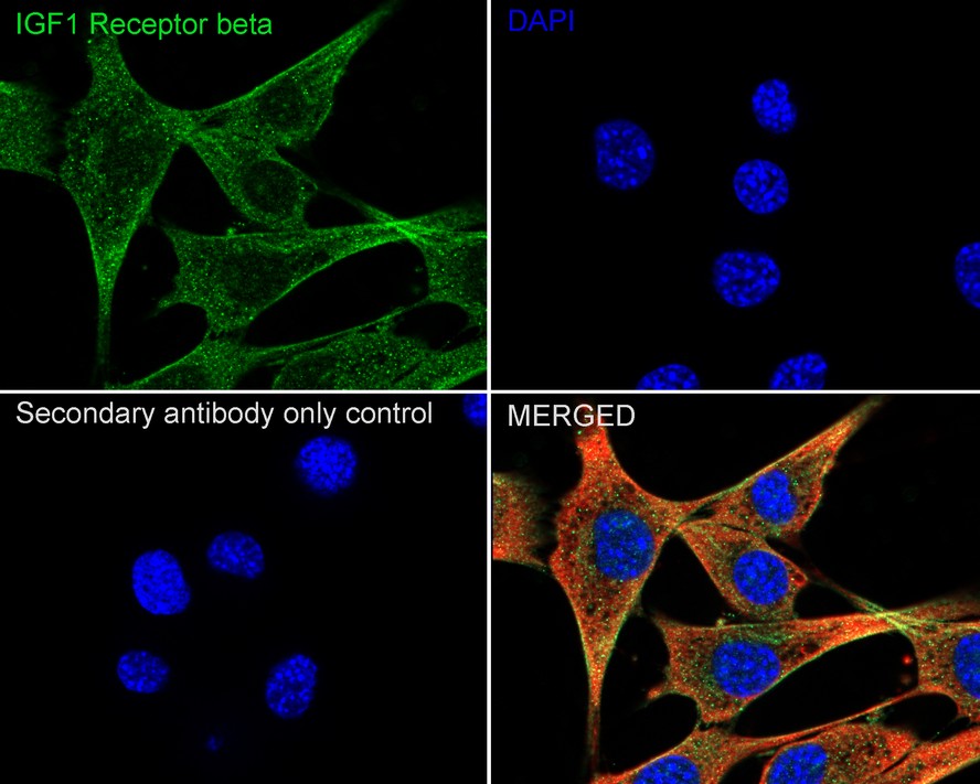
Immunocytochemistry analysis of NIH/3T3 cells labeling IGF1 Receptor beta with Rabbit anti-IGF1 Receptor beta antibody at 1/100 dilution. Cells were fixed in 4% paraformaldehyde for 15 minutes at room temperature, permeabilized with 0.1% Triton X-100 in PBS for 15 minutes at room temperature, then blocked with 1% BSA in 10% negative goat serum for 1 hour at room temperature. Cells were then incubated with Rabbit anti-IGF1 Receptor beta antibody at 1/100 dilution in 1% BSA in PBST overnight at 4 ℃. Goat Anti-Rabbit IgG H&L (488) was used as the secondary antibody at 1/1,000 dilution. PBS instead of the primary antibody was used as the secondary antibody only control. Nuclear DNA was labelled in blue with DAPI. Beta tubulin (red) was stained at 1/100 dilution overnight at +4℃. Goat Anti-Mouse IgG H&L (594) was used as the secondary antibody at 1/1,000 dilution.| Product Name | IGF1 Receptor beta Recombinant Rabbit Monoclonal Antibody |
|---|---|
| Antibody Type | Primary Antibodies |
| Immunogen | Recombinant protein within human IGF1 Receptor aa 701-935. |
| Clonality | monoclonal |
|---|---|
| Isotype | IgG |
| Host Species | Rabbit |
| Tested Applications | ICC/IFWB |
| WB:1:2000 ICC/IF:1:100 |
|
| Species Reactivity | HumanMouseRat |
| Concentration | 1mg/ml |
| Purification | Protein A |
| Gene Symbol | IGF1R |
|---|---|
| Gene Synonyms | IGFR CD221 IGFIR JTK13 |
| Gene Full Name | insulin like growth factor 1 receptor |
| Gene Summary | This receptor binds insulin-like growth factor with a high affinity. It has tyrosine kinase activity. The insulin-like growth factor I receptor plays a critical role in transformation events. Cleavage of the precursor generates alpha and beta subunits. It is highly overexpressed in most malignant tissues where it functions as an anti-apoptotic agent by enhancing cell survival. Alternatively spliced transcript variants encoding distinct isoforms have been found for this gene. [provided by RefSeq, May 2014] |
| Molecular Weight(MW) | 155kDa(Observed band size: 95kDa) |
| Cellular Localization | Cell membrane. |
WB
Western blot analysis of IGF1 Receptor beta on different lysates with Rabbit anti-IGF1 Receptor beta antibody at 1/2,000 dilution. Lane 1: HEK-293 cell lysate (20 µg/Lane), Lane 2: MCF7 cell lysate (20 µg/Lane), Lane 3: MDA-MB-231 cell lysate (low expression) (20 µg/Lane), Lane 4: A431 cell lysate (20 µg/Lane), Lane 5: HepG2 cell lysate (20 µg/Lane), Lane 6: A549 cell lysate (20 µg/Lane), Lane 7: HeLa cell lysate (20 µg/Lane), Lane 8: HUVEC cell lysate (20 µg/Lane), Lane 9: NIH/3T3 cell lysate (20 µg/Lane), Lane 10: C2C12 cell lysate (20 µg/Lane), Lane 11: C6 cell lysate (20 µg/Lane), Lane 12: Mouse brain tissue lysate (40 µg/Lane), Lane 13: Rat brain tissue lysate (40 µg/Lane), Exposure time: 3 minutes; 4-20% SDS-PAGE gel. Proteins were transferred to a PVDF membrane and blocked with 5% NFDM/TBST for 1 hour at room temperature. The primary antibody at 1/2,000 dilution was used in 5% NFDM/TBST at 4℃ overnight. Goat Anti-Rabbit IgG - HRP Secondary Antibody at 1/50,000 dilution was used for 1 hour at room temperature.
ICC/IF
Immunocytochemistry analysis of NIH/3T3 cells labeling IGF1 Receptor beta with Rabbit anti-IGF1 Receptor beta antibody at 1/100 dilution. Cells were fixed in 4% paraformaldehyde for 15 minutes at room temperature, permeabilized with 0.1% Triton X-100 in PBS for 15 minutes at room temperature, then blocked with 1% BSA in 10% negative goat serum for 1 hour at room temperature. Cells were then incubated with Rabbit anti-IGF1 Receptor beta antibody at 1/100 dilution in 1% BSA in PBST overnight at 4 ℃. Goat Anti-Rabbit IgG H&L (488) was used as the secondary antibody at 1/1,000 dilution. PBS instead of the primary antibody was used as the secondary antibody only control. Nuclear DNA was labelled in blue with DAPI. Beta tubulin (red) was stained at 1/100 dilution overnight at +4℃. Goat Anti-Mouse IgG H&L (594) was used as the secondary antibody at 1/1,000 dilution.| Application Notes | WB:1:2000 ICC/IF:1:100 |
|---|
| Form | Liquid |
|---|---|
| Storage Instructions | Store at +4℃ after thawing. Aliquot store at -20℃. Avoid repeated freeze / thaw cycles. |
| Storage Buffer | 1*TBS (pH7.4), 0.05% BSA, 40% Glycerol. Preservative: 0.05% Sodium Azide. |


 Data sheet for OM643992