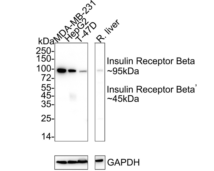
WB
Western blot analysis of Insulin Receptor Beta on different lysates with Rabbit anti-Insulin Receptor Beta antibody at 1/1,000 dilution. Lane 1: MDA-MB-231 cell lysate, Lane 2: HepG2 cell lysate, Lane 3: T-47D cell lysate, Lane 4: Rat liver tissue lysate, Cell lysates/proteins at 20 µg/Lane. Tissue lysates/proteins at 20 µg/Lane. Exposure time: 1 minutes 2 seconds; 4-20% SDS-PAGE gel. Proteins were transferred to a PVDF membrane and blocked with 5% NFDM/TBST for 1 hour at room temperature. The primary antibody at 1/1,000 dilution was used in 5% NFDM/TBST at 4℃ overnight. Goat Anti-Rabbit IgG - HRP Secondary Antibody at 1/50,000 dilution was used for 1 hour at room temperature.IHC
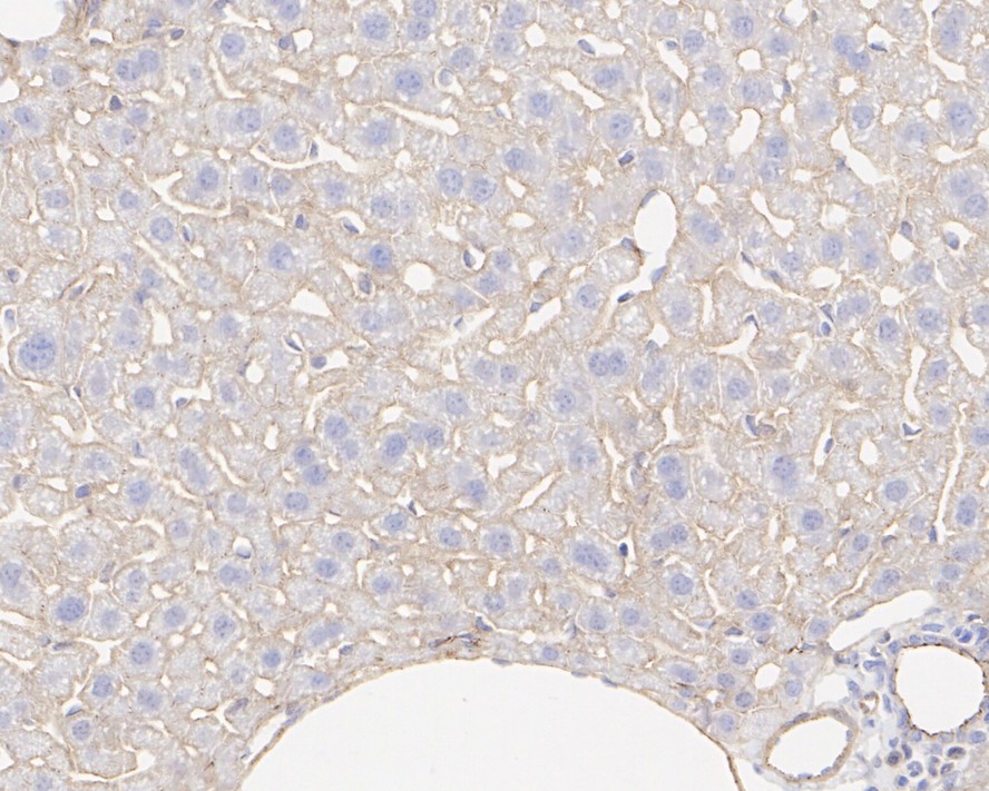
Immunohistochemical analysis of paraffin-embedded mouse liver tissue with Rabbit anti-Insulin Receptor Beta antibody at 1/1,000 dilution. The section was pre-treated using heat mediated antigen retrieval with Tris-EDTA buffer (pH 9.0) for 20 minutes. The tissues were blocked in 1% BSA for 20 minutes at room temperature, washed with ddH2O and PBS, and then probed with the primary antibody at 1/1,000 dilution for 1 hour at room temperature. The detection was performed using an HRP conjugated compact polymer system. DAB was used as the chromogen. Tissues were counterstained with hematoxylin and mounted with DPX.ICC/IF
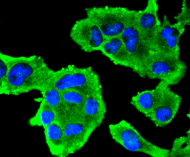
ICC staining of Insulin Receptor Beta in RH-35 cells (green). Formalin fixed cells were permeabilized with 0.1% Triton X-100 in TBS for 10 minutes at room temperature and blocked with 1% Blocker BSA for 15 minutes at room temperature. Cells were probed with the primary antibody (1/50) for 1 hour at room temperature, washed with PBS. Alexa Fluor®488 Goat anti-Rabbit IgG was used as the secondary antibody at 1/1,000 dilution. The nuclear counter stain is DAPI (blue).| Product Name | Insulin Receptor Beta Recombinant Rabbit Monoclonal Antibody |
|---|---|
| Antibody Type | Primary Antibodies |
| Immunogen | Synthetic peptide within human Insulin receptor aa 930-970. |
| Clonality | monoclonal |
|---|---|
| Isotype | IgG |
| Host Species | Rabbit |
| Tested Applications | ICC/IFIHCWB |
| WB:1:500-1:2000 IHC:1:1000 ICC/IF:1:100-1:500 |
|
| Species Reactivity | HumanMouseRat |
| Concentration | 1mg/ml |
| Purification | Protein A |
| Gene Symbol | INSR |
|---|---|
| Gene Synonyms | HHF5 CD220 |
| Gene Full Name | insulin receptor |
| Gene Summary | This gene encodes a member of the receptor tyrosine kinase family of proteins. The encoded preproprotein is proteolytically processed to generate alpha and beta subunits that form a heterotetrameric receptor. Binding of insulin or other ligands to this receptor activates the insulin signaling pathway, which regulates glucose uptake and release, as well as the synthesis and storage of carbohydrates, lipids and protein. Mutations in this gene underlie the inherited severe insulin resistance syndromes including type A insulin resistance syndrome, Donohue syndrome and Rabson-Mendenhall syndrome. Alternative splicing results in multiple transcript variants. [provided by RefSeq, Oct 2015] |
| Molecular Weight(MW) | 156kDa(Observed band size: 95kDa) |
| Cellular Localization | Cell membrane. |
WB
Western blot analysis of Insulin Receptor Beta on different lysates with Rabbit anti-Insulin Receptor Beta antibody at 1/1,000 dilution. Lane 1: MDA-MB-231 cell lysate, Lane 2: HepG2 cell lysate, Lane 3: T-47D cell lysate, Lane 4: Rat liver tissue lysate, Cell lysates/proteins at 20 µg/Lane. Tissue lysates/proteins at 20 µg/Lane. Exposure time: 1 minutes 2 seconds; 4-20% SDS-PAGE gel. Proteins were transferred to a PVDF membrane and blocked with 5% NFDM/TBST for 1 hour at room temperature. The primary antibody at 1/1,000 dilution was used in 5% NFDM/TBST at 4℃ overnight. Goat Anti-Rabbit IgG - HRP Secondary Antibody at 1/50,000 dilution was used for 1 hour at room temperature.
IHC
Immunohistochemical analysis of paraffin-embedded mouse liver tissue with Rabbit anti-Insulin Receptor Beta antibody at 1/1,000 dilution. The section was pre-treated using heat mediated antigen retrieval with Tris-EDTA buffer (pH 9.0) for 20 minutes. The tissues were blocked in 1% BSA for 20 minutes at room temperature, washed with ddH2O and PBS, and then probed with the primary antibody at 1/1,000 dilution for 1 hour at room temperature. The detection was performed using an HRP conjugated compact polymer system. DAB was used as the chromogen. Tissues were counterstained with hematoxylin and mounted with DPX.
ICC/IF
ICC staining of Insulin Receptor Beta in RH-35 cells (green). Formalin fixed cells were permeabilized with 0.1% Triton X-100 in TBS for 10 minutes at room temperature and blocked with 1% Blocker BSA for 15 minutes at room temperature. Cells were probed with the primary antibody (1/50) for 1 hour at room temperature, washed with PBS. Alexa Fluor®488 Goat anti-Rabbit IgG was used as the secondary antibody at 1/1,000 dilution. The nuclear counter stain is DAPI (blue).| Application Notes | WB:1:500-1:2000 IHC:1:1000 ICC/IF:1:100-1:500 |
|---|
| Form | Liquid |
|---|---|
| Storage Instructions | Store at +4℃ after thawing. Aliquot store at -20℃. Avoid repeated freeze / thaw cycles. |
| Storage Buffer | 1*TBS (pH7.4), 0.05% BSA, 40% Glycerol. Preservative: 0.05% Sodium Azide. |


 Data sheet for OM643994