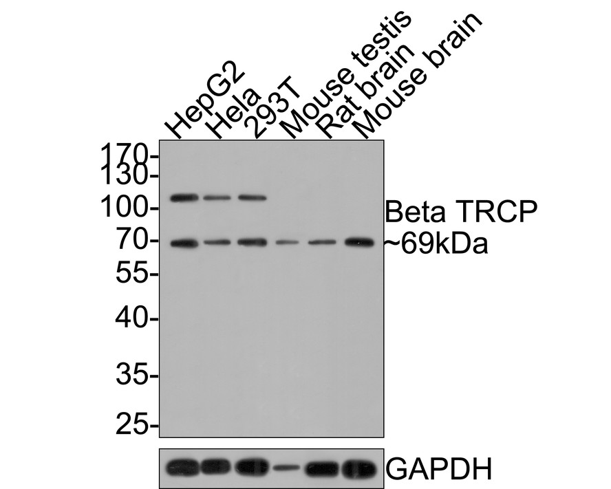
WB
Western blot analysis of Beta TRCP on different lysates with Rabbit anti-Beta TRCP antibody at 1/1,000 dilution. Lane 1: HepG2 cell lysate (10 µg/Lane), Lane 2: Hela cell lysate (10 µg/Lane), Lane 3: 293T cell lysate (10 µg/Lane), Lane 4: Mouse testis tissue lysate (20 µg/Lane), Lane 5: Rat brain tissue lysate (20 µg/Lane), Lane 6: Mouse brain tissue lysate (20 µg/Lane), Exposure time: 2 minutes; 10% SDS-PAGE gel. Proteins were transferred to a PVDF membrane and blocked with 5% NFDM/TBST for 1 hour at room temperature. The primary antibody at 1/1,000 dilution was used in 5% NFDM/TBST at room temperature for 2 hours. Goat Anti-Rabbit IgG - HRP Secondary Antibody at 1:300,000 dilution was used for 1 hour at room temperature.IHC
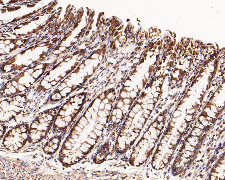
Immunohistochemical analysis of paraffin-embedded human colon tissue with Rabbit anti-Beta TRCP antibody at 1/1,000 dilution. The section was pre-treated using heat mediated antigen retrieval with sodium citrate buffer (pH 6.0) for 2 minutes. The tissues were blocked in 1% BSA for 20 minutes at room temperature, washed with ddH2O and PBS, and then probed with the primary antibody at 1/1,000 dilution for 1 hour at room temperature. The detection was performed using an HRP conjugated compact polymer system. DAB was used as the chromogen. Tissues were counterstained with hematoxylin and mounted with DPX.ICC/IF
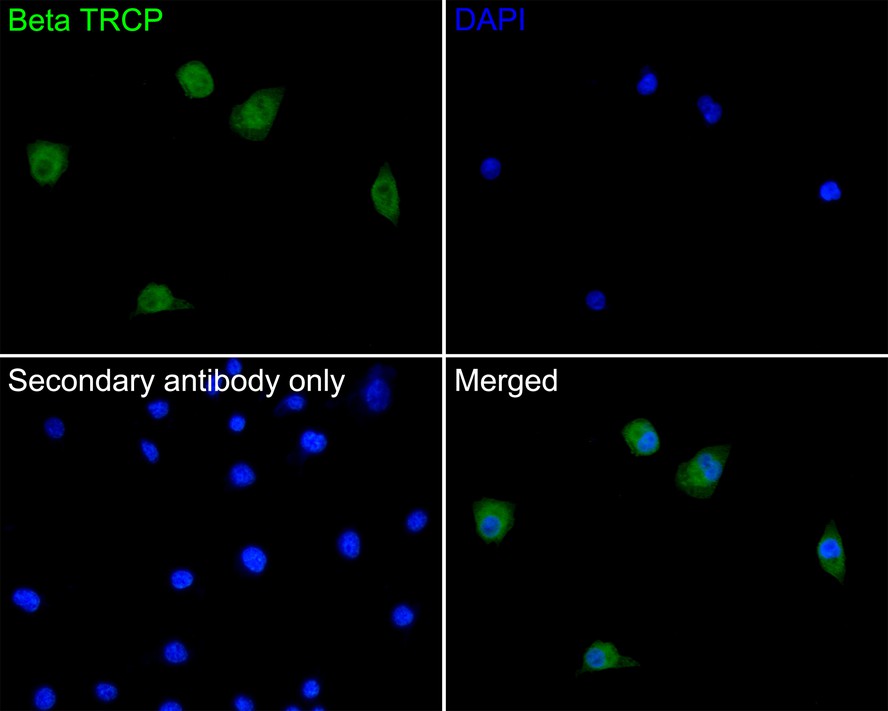
Immunocytochemistry analysis of A549 cells labeling Beta TRCP with Rabbit anti-Beta TRCP antibody at 1/100 dilution. Cells were fixed in 4% paraformaldehyde for 10 minutes at 37 ℃, permeabilized with 0.05% Triton X-100 in PBS for 20 minutes, and then blocked with 2% negative goat serum for 30 minutes at room temperature. Cells were then incubated with Rabbit anti-Beta TRCP antibody at 1/100 dilution in 2% negative goat serum overnight at 4 ℃. Goat Anti-Rabbit IgG H&L (488) was used as the secondary antibody at 1/1,000 dilution. PBS instead of the primary antibody was used as the secondary antibody only control. Nuclear DNA was labelled in blue with DAPI.| Product Name | Beta TRCP Rabbit Polyclonal Antibody |
|---|---|
| Antibody Type | Primary Antibodies |
| Immunogen | Recombinant protein within Human Beta TRCP aa 1-200 / 605. |
| Clonality | polyclonal |
|---|---|
| Isotype | IgG |
| Host Species | Rabbit |
| Tested Applications | ICC/IFIHCWB |
| WB:1:1000 IHC:1:1000-1:2000 ICC/IF:1:100 |
|
| Species Reactivity | HumanMouseRat |
| Concentration | 1mg/ml |
| Purification | Protein A |
| Gene Symbol | BTRC |
|---|---|
| Gene Synonyms | FWD1 FBW1A FBXW1 bTrCP FBXW1A bTrCP1 betaTrCP BETA-TRCP |
| Gene Full Name | beta-transducin repeat containing E3 ubiquitin protein ligase |
| Gene Summary | This gene encodes a member of the F-box protein family which is characterized by an approximately 40 amino acid motif, the F-box. The F-box proteins constitute one of the four subunits of ubiquitin protein ligase complex called SCFs (SKP1-cullin-F-box), which function in phosphorylation-dependent ubiquitination. The F-box proteins are divided into 3 classes: Fbws containing WD-40 domains, Fbls containing leucine-rich repeats, and Fbxs containing either different protein-protein interaction modules or no recognizable motifs. The protein encoded by this gene belongs to the Fbws class; in addition to an F-box, this protein contains multiple WD-40 repeats. The encoded protein mediates degradation of CD4 via its interaction with HIV-1 Vpu. It has also been shown to ubiquitinate phosphorylated NFKBIA (nuclear factor of kappa light polypeptide gene enhancer in B-cells inhibitor, alpha), targeting it for degradation and thus activating nuclear factor kappa-B. Alternatively spliced transcript variants have been described. A related pseudogene exists in chromosome 6. [provided by RefSeq, Mar 2012] |
| Molecular Weight(MW) | 69/110kDa |
| Cellular Localization | Cytoplasm, Nucleus. |
WB
Western blot analysis of Beta TRCP on different lysates with Rabbit anti-Beta TRCP antibody at 1/1,000 dilution. Lane 1: HepG2 cell lysate (10 µg/Lane), Lane 2: Hela cell lysate (10 µg/Lane), Lane 3: 293T cell lysate (10 µg/Lane), Lane 4: Mouse testis tissue lysate (20 µg/Lane), Lane 5: Rat brain tissue lysate (20 µg/Lane), Lane 6: Mouse brain tissue lysate (20 µg/Lane), Exposure time: 2 minutes; 10% SDS-PAGE gel. Proteins were transferred to a PVDF membrane and blocked with 5% NFDM/TBST for 1 hour at room temperature. The primary antibody at 1/1,000 dilution was used in 5% NFDM/TBST at room temperature for 2 hours. Goat Anti-Rabbit IgG - HRP Secondary Antibody at 1:300,000 dilution was used for 1 hour at room temperature.
IHC
Immunohistochemical analysis of paraffin-embedded human colon tissue with Rabbit anti-Beta TRCP antibody at 1/1,000 dilution. The section was pre-treated using heat mediated antigen retrieval with sodium citrate buffer (pH 6.0) for 2 minutes. The tissues were blocked in 1% BSA for 20 minutes at room temperature, washed with ddH2O and PBS, and then probed with the primary antibody at 1/1,000 dilution for 1 hour at room temperature. The detection was performed using an HRP conjugated compact polymer system. DAB was used as the chromogen. Tissues were counterstained with hematoxylin and mounted with DPX.
ICC/IF
Immunocytochemistry analysis of A549 cells labeling Beta TRCP with Rabbit anti-Beta TRCP antibody at 1/100 dilution. Cells were fixed in 4% paraformaldehyde for 10 minutes at 37 ℃, permeabilized with 0.05% Triton X-100 in PBS for 20 minutes, and then blocked with 2% negative goat serum for 30 minutes at room temperature. Cells were then incubated with Rabbit anti-Beta TRCP antibody at 1/100 dilution in 2% negative goat serum overnight at 4 ℃. Goat Anti-Rabbit IgG H&L (488) was used as the secondary antibody at 1/1,000 dilution. PBS instead of the primary antibody was used as the secondary antibody only control. Nuclear DNA was labelled in blue with DAPI.| Application Notes | WB:1:1000 IHC:1:1000-1:2000 ICC/IF:1:100 |
|---|
| Form | Liquid |
|---|---|
| Storage Instructions | Store at +4℃ after thawing. Aliquot store at -20℃. Avoid repeated freeze / thaw cycles. |
| Storage Buffer | 1*TBS (pH7.4), 0.05% BSA, 40% Glycerol. Preservative: 0.05% Sodium Azide. |


 Data sheet for OM643998