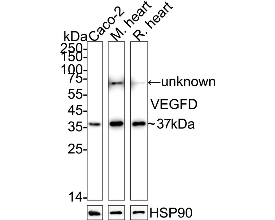
WB
Western blot analysis of VEGFD on different lysates with Rabbit anti-VEGFD antibody at 1/2,000 dilution. Lane 1: Caco-2 cell lysate, Lane 2: Mouse heart tissue lysate, Lane 3: Rat heart tissue lysate, Lysates/proteins at 20 µg/Lane. Exposure time: 20 seconds; 4-20% SDS-PAGE gel. Proteins were transferred to a PVDF membrane and blocked with 5% NFDM/TBST for 1 hour at room temperature. The primary antibody at 1/2,000 dilution was used in 5% NFDM/TBST at 4℃ overnight. Goat Anti-Rabbit IgG - HRP Secondary Antibody at 1/50,000 dilution was used for 1 hour at room temperature.IHC
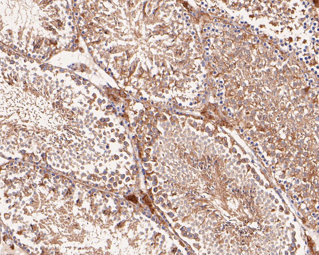
Immunohistochemical analysis of paraffin-embedded mouse testis tissue with Rabbit anti-VEGFD antibody at 1/50 dilution. The section was pre-treated using heat mediated antigen retrieval with Tris-EDTA buffer (pH 8.0-8.4) for 20 minutes. The tissues were blocked in 1% BSA for 20 minutes at room temperature, washed with ddH2O and PBS, and then probed with the primary antibody at 1/50 dilution for 0.5 hour at room temperature. The detection was performed using an HRP conjugated compact polymer system. DAB was used as the chromogen. Tissues were counterstained with hematoxylin and mounted with DPX.ICC/IF
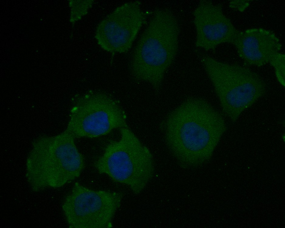
Immunocytochemistry analysis of HUVEC cells labeling VEGFD with Rabbit anti-VEGFD antibody at 1/100 dilution. Cells were fixed in 4% paraformaldehyde for 10 minutes at 37 ℃, permeabilized with 0.05% Triton X-100 in PBS for 20 minutes, and then blocked with 2% negative goat serum for 30 minutes at room temperature. Cells were then incubated with Rabbit anti-VEGFD antibody at 1/100 dilution in 2% negative goat serum overnight at 4 ℃.Alexa Fluor®488 Goat anti-Rabbit IgG was used as the secondary antibody at 1/1,000 dilution.Nuclear DNA was labelled in blue with DAPI.FC
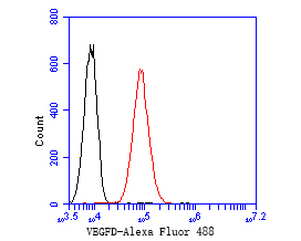
Flow cytometric analysis of VEGFD was done on A549 cells. The cells were fixed, permeabilized and stained with the primary antibody (1/50) (red). After incubation of the primary antibody at room temperature for an hour, the cells were stained with a Alexa Fluor 488-conjugated Goat anti-Rabbit IgG Secondary antibody at 1/1000 dilution for 30 minutes.Unlabelled sample was used as a control (cells without incubation with primary antibody; black).| Product Name | VEGFD Recombinant Rabbit Monoclonal Antibody |
|---|---|
| Antibody Type | Primary Antibodies |
| Immunogen | Recombinant protein within Human VEGFD aa 181-302 / 354. |
| Clonality | monoclonal |
|---|---|
| Isotype | IgG |
| Host Species | Rabbit |
| Tested Applications | FCICC/IFIHCWB |
| WB:1:2000 IHC:1:50-1:200 ICC/IF:1:50-1:100 FC:1:50-1:100 |
|
| Species Reactivity | HumanMouseRat |
| Concentration | 1mg/ml |
| Purification | Protein A |
| Gene Symbol | VEGFD |
|---|---|
| Gene Synonyms | FIGF VEGF-D |
| Gene Full Name | vascular endothelial growth factor D |
| Gene Summary | The protein encoded by this gene is a member of the platelet-derived growth factor/vascular endothelial growth factor (PDGF/VEGF) family and is active in angiogenesis, lymphangiogenesis, and endothelial cell growth. This secreted protein undergoes a complex proteolytic maturation, generating multiple processed forms which bind and activate VEGFR-2 and VEGFR-3 receptors. This protein is structurally and functionally similar to vascular endothelial growth factor C. Read-through transcription has been observed between this locus and the upstream PIR (GeneID 8544) locus. [provided by RefSeq, Feb 2011] |
| Molecular Weight(MW) | 40kDa(Observed band size: 37kDa) |
| Cellular Localization | Secreted. |
WB
Western blot analysis of VEGFD on different lysates with Rabbit anti-VEGFD antibody at 1/2,000 dilution. Lane 1: Caco-2 cell lysate, Lane 2: Mouse heart tissue lysate, Lane 3: Rat heart tissue lysate, Lysates/proteins at 20 µg/Lane. Exposure time: 20 seconds; 4-20% SDS-PAGE gel. Proteins were transferred to a PVDF membrane and blocked with 5% NFDM/TBST for 1 hour at room temperature. The primary antibody at 1/2,000 dilution was used in 5% NFDM/TBST at 4℃ overnight. Goat Anti-Rabbit IgG - HRP Secondary Antibody at 1/50,000 dilution was used for 1 hour at room temperature.
IHC
Immunohistochemical analysis of paraffin-embedded mouse testis tissue with Rabbit anti-VEGFD antibody at 1/50 dilution. The section was pre-treated using heat mediated antigen retrieval with Tris-EDTA buffer (pH 8.0-8.4) for 20 minutes. The tissues were blocked in 1% BSA for 20 minutes at room temperature, washed with ddH2O and PBS, and then probed with the primary antibody at 1/50 dilution for 0.5 hour at room temperature. The detection was performed using an HRP conjugated compact polymer system. DAB was used as the chromogen. Tissues were counterstained with hematoxylin and mounted with DPX.
ICC/IF
Immunocytochemistry analysis of HUVEC cells labeling VEGFD with Rabbit anti-VEGFD antibody at 1/100 dilution. Cells were fixed in 4% paraformaldehyde for 10 minutes at 37 ℃, permeabilized with 0.05% Triton X-100 in PBS for 20 minutes, and then blocked with 2% negative goat serum for 30 minutes at room temperature. Cells were then incubated with Rabbit anti-VEGFD antibody at 1/100 dilution in 2% negative goat serum overnight at 4 ℃.Alexa Fluor®488 Goat anti-Rabbit IgG was used as the secondary antibody at 1/1,000 dilution.Nuclear DNA was labelled in blue with DAPI.
FC
Flow cytometric analysis of VEGFD was done on A549 cells. The cells were fixed, permeabilized and stained with the primary antibody (1/50) (red). After incubation of the primary antibody at room temperature for an hour, the cells were stained with a Alexa Fluor 488-conjugated Goat anti-Rabbit IgG Secondary antibody at 1/1000 dilution for 30 minutes.Unlabelled sample was used as a control (cells without incubation with primary antibody; black).| Application Notes | WB:1:2000 IHC:1:50-1:200 ICC/IF:1:50-1:100 FC:1:50-1:100 |
|---|
| Form | Liquid |
|---|---|
| Storage Instructions | Store at +4℃ after thawing. Aliquot store at -20℃. Avoid repeated freeze / thaw cycles. |
| Storage Buffer | 1*TBS (pH7.4), 0.05% BSA, 40% Glycerol. Preservative: 0.05% Sodium Azide. |


 Data sheet for OM644004