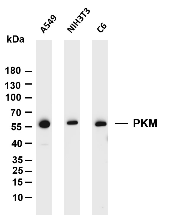
WB
Various whole cell lysates were separated by 4-20% SDS-PAGE, and the membrane was blotted with anti-PKM antibody. The HRP conjugated Goat anti-Rabbit IgG(H + L) antibody was used to detect the antibody. Lane 1: A549, Lane 2: NIH3T3, Lane 3: C6.IHC
Mouse lung was stained with Anti-PKM rabbit antibody.ICC/IF
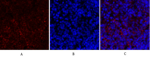
Immunofluorescence analysis of rat-spleen tissue. 1,PKM2 Antibody(red) was diluted at 1:200(4°C,overnight). 2, Cy3 labled Secondary antibody was diluted at 1:300(room temperature, 50min).3, Picture B: DAPI(blue) 10min. Picture A:Target. Picture B: DAPI. Picture C: merge of A+B.| Product Name | PKM Rabbit mAb |
|---|---|
| Antibody Type | Primary Antibodies |
| Clonality | monoclonal |
|---|---|
| Isotype | IgG |
| Host Species | Rabbit |
| Tested Applications | ICC/IFIHCWB |
| WB:1:1000-1:5000 IHC:1:200-1:1000 ICC/IF:1:200-1:1000 |
|
| Species Reactivity | HumanMouseRat |
| Concentration | 1mg/ml |
| Purification | Protein A |
| Gene Symbol | PKM |
|---|---|
| Gene Synonyms | PK3 TCB p58 OIP3 PKM2 CTHBP THBP1 HEL-S-30 |
| Gene Full Name | pyruvate kinase M1/2 |
| Gene Summary | This gene encodes a protein involved in glycolysis. The encoded protein is a pyruvate kinase that catalyzes the transfer of a phosphoryl group from phosphoenolpyruvate to ADP, generating ATP and pyruvate. This protein has been shown to interact with thyroid hormone and may mediate cellular metabolic effects induced by thyroid hormones. This protein has been found to bind Opa protein, a bacterial outer membrane protein involved in gonococcal adherence to and invasion of human cells, suggesting a role of this protein in bacterial pathogenesis. Several alternatively spliced transcript variants encoding a few distinct isoforms have been reported. [provided by RefSeq, May 2011] |
| Molecular Weight(MW) | 57kDa |
| Cellular Localization | Cytoplasm,Nucleus. |
WB
Various whole cell lysates were separated by 4-20% SDS-PAGE, and the membrane was blotted with anti-PKM antibody. The HRP conjugated Goat anti-Rabbit IgG(H + L) antibody was used to detect the antibody. Lane 1: A549, Lane 2: NIH3T3, Lane 3: C6.
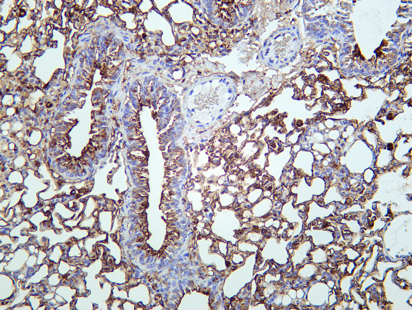
IHC
Mouse lung was stained with Anti-PKM rabbit antibody.
ICC/IF
Immunofluorescence analysis of rat-spleen tissue. 1,PKM2 Antibody(red) was diluted at 1:200(4°C,overnight). 2, Cy3 labled Secondary antibody was diluted at 1:300(room temperature, 50min).3, Picture B: DAPI(blue) 10min. Picture A:Target. Picture B: DAPI. Picture C: merge of A+B.| Application Notes | WB:1:1000-1:5000 IHC:1:200-1:1000 ICC/IF:1:200-1:1000 |
|---|
| Form | Liquid |
|---|---|
| Storage Instructions | -15°C to -25°C/1 year(Do not lower than -25°C) |
| Storage Buffer | PBS, 50% glycerol, 0.05% Proclin 300, 0.05%BSA |


 Data sheet for OM644038