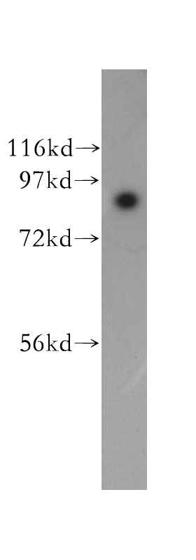
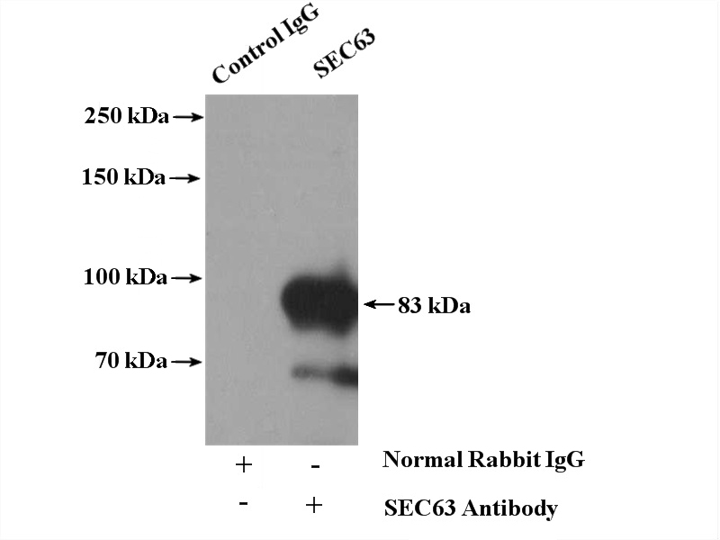
WB
A431 cells were subjected to SDS PAGE followed by western blot with SEC63 antibody at dilution of 1:1000 incubated at room temperature for 1.5 hours.IHC
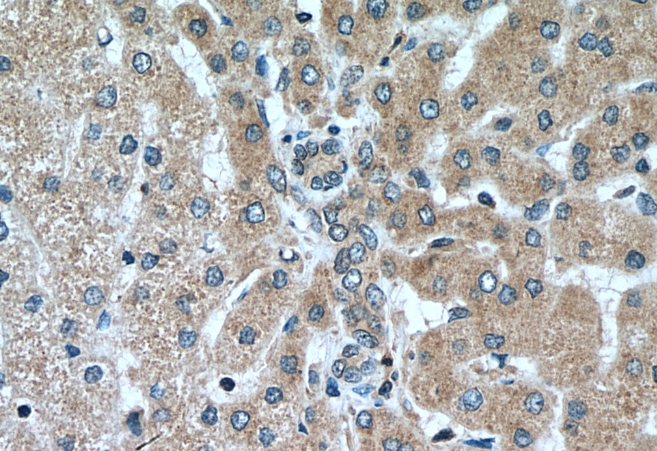
Immunohistochemical analysis of paraffin-embedded human liver tissue slide using SEC63 antibody at dilution of 1:200 (under 40x lens). Heat mediated antigen retrieval with Tris-EDTA buffer (pH 9.0).ICC/IF
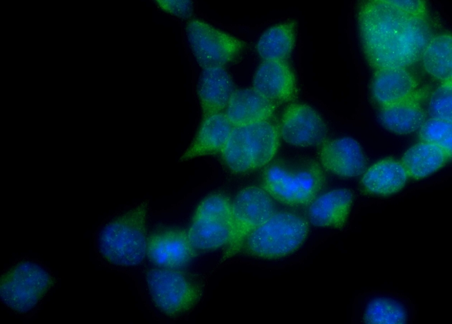
Immunofluorescent analysis of (-20°C Ethanol) fixed HEK-293 cells using SEC63 antibody at dilution of 1:200 and Omnimabs® 488 Goat Anti-Rabbit IgG(H&L).IF-P
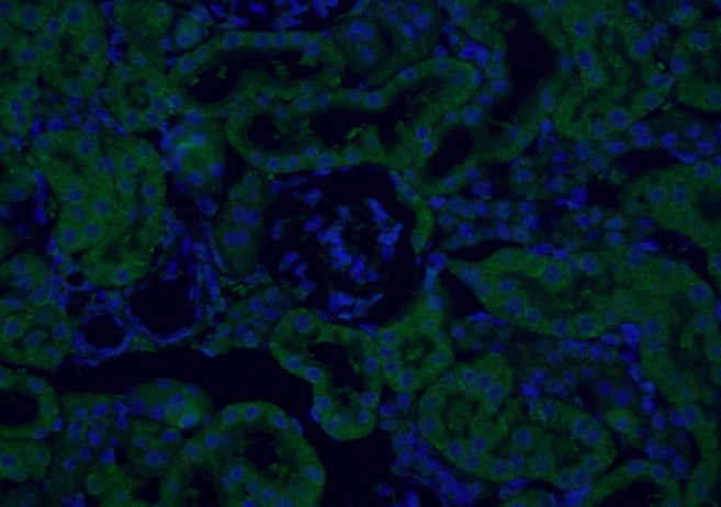
Immunofluorescent analysis of (4% PFA) fixed mouse kidney tissue using SEC63 antibody at dilution of 1:50 and Alexa Fluor 488-Conjugated Goat Anti-Rabbit IgG(H+L).IP
IP result of anti-SEC63 (IP:4ug; Detection:1:300) with A431 cells lysate 2.4mg.| Product Name | SEC63 Polyclonal antibody |
|---|---|
| Antibody Type | Primary Antibodies |
| Immunogen | Recombinant human SEC63 protein |
| Clonality | polyclonal |
|---|---|
| Isotype | IgG |
| Host Species | Rabbit |
| Tested Applications | ICC/IFIF-PIHCIPWB |
| WB:1:1000-1:4000 IHC:1:50-1:500 ICC/IF:1:50-1:500 IF-P:1:50-1:500 IP:0.5-4.0 ug for 1.0-3.0 mg of total protein lysate |
|
| Species Reactivity | HumanMouseRat |
| Concentration | 0.8mg/ml |
| Purification | Affinity purified |
| Gene Symbol | SEC63 |
|---|---|
| Gene Synonyms | ERdj2 PCLD2 SEC63L DNAJC23 PRO2507 |
| Gene Full Name | SEC63 homolog, protein translocation regulator |
| Gene Summary | The Sec61 complex is the central component of the protein translocation apparatus of the endoplasmic reticulum (ER) membrane. The protein encoded by this gene and SEC62 protein are found to be associated with ribosome-free SEC61 complex. It is speculated that Sec61-Sec62-Sec63 may perform post-translational protein translocation into the ER. The Sec61-Sec62-Sec63 complex might also perform the backward transport of ER proteins that are subject to the ubiquitin-proteasome-dependent degradation pathway. The encoded protein is an integral membrane protein located in the rough ER. [provided by RefSeq, Jul 2008] |
| Molecular Weight(MW) | 83-90kDa |
| Cellular Localization | Endoplasmic reticulum membrane. |
WB
A431 cells were subjected to SDS PAGE followed by western blot with SEC63 antibody at dilution of 1:1000 incubated at room temperature for 1.5 hours.
IHC
Immunohistochemical analysis of paraffin-embedded human liver tissue slide using SEC63 antibody at dilution of 1:200 (under 40x lens). Heat mediated antigen retrieval with Tris-EDTA buffer (pH 9.0).
ICC/IF
Immunofluorescent analysis of (-20°C Ethanol) fixed HEK-293 cells using SEC63 antibody at dilution of 1:200 and Omnimabs® 488 Goat Anti-Rabbit IgG(H&L).
IF-P
Immunofluorescent analysis of (4% PFA) fixed mouse kidney tissue using SEC63 antibody at dilution of 1:50 and Alexa Fluor 488-Conjugated Goat Anti-Rabbit IgG(H+L).
IP
IP result of anti-SEC63 (IP:4ug; Detection:1:300) with A431 cells lysate 2.4mg.| Application Notes | WB:1:1000-1:4000 IHC:1:50-1:500 ICC/IF:1:50-1:500 IF-P:1:50-1:500 IP:0.5-4.0 ug for 1.0-3.0 mg of total protein lysate |
|---|
| Form | Liquid |
|---|---|
| Storage Instructions | Store at -20°C. Stable for one year after shipment. |
| Storage Buffer | PBS with 0.02% sodium azide and 50% glycerol pH 7.3. |


 Data sheet for OM644094