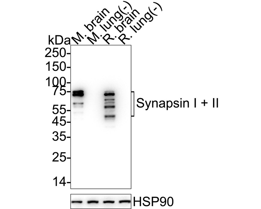
WB
Western blot analysis of Synapsin I + II on different lysates with Rabbit anti-Synapsin I + II antibody at 1/5,000 dilution. Lane 1: Mouse brain tissue lysate, Lane 2: Mouse lung tissue lysate (negative), Lane 3: Rat brain tissue lysate, Lane 4: Rat lung tissue lysate (negative), Lysates/proteins at 20 µg/Lane. Exposure time: 6 seconds; 4-20% SDS-PAGE gel. Proteins were transferred to a PVDF membrane and blocked with 5% NFDM/TBST for 1 hour at room temperature. The primary antibody at 1/5,000 dilution was used in 5% NFDM/TBST at 4℃ overnight. Goat Anti-Rabbit IgG - HRP Secondary Antibody at 1/50,000 dilution was used for 1 hour at room temperature.IHC
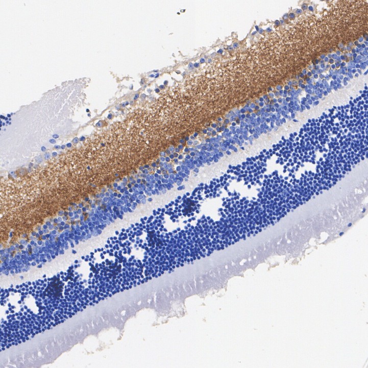
Immunohistochemical analysis of paraffin-embedded mouse retina tissue with Rabbit anti-Synapsin I + II antibody at 1/200 dilution. The section was pre-treated using heat mediated antigen retrieval with Tris-EDTA buffer (pH 9.0) for 20 minutes. The tissues were blocked in 1% BSA for 20 minutes at room temperature, washed with ddH2O and PBS, and then probed with the primary antibody at 1/200 dilution for 1 hour at room temperature. The detection was performed using an HRP conjugated compact polymer system. DAB was used as the chromogen. Tissues were counterstained with hematoxylin and mounted with DPX.ICC/IF
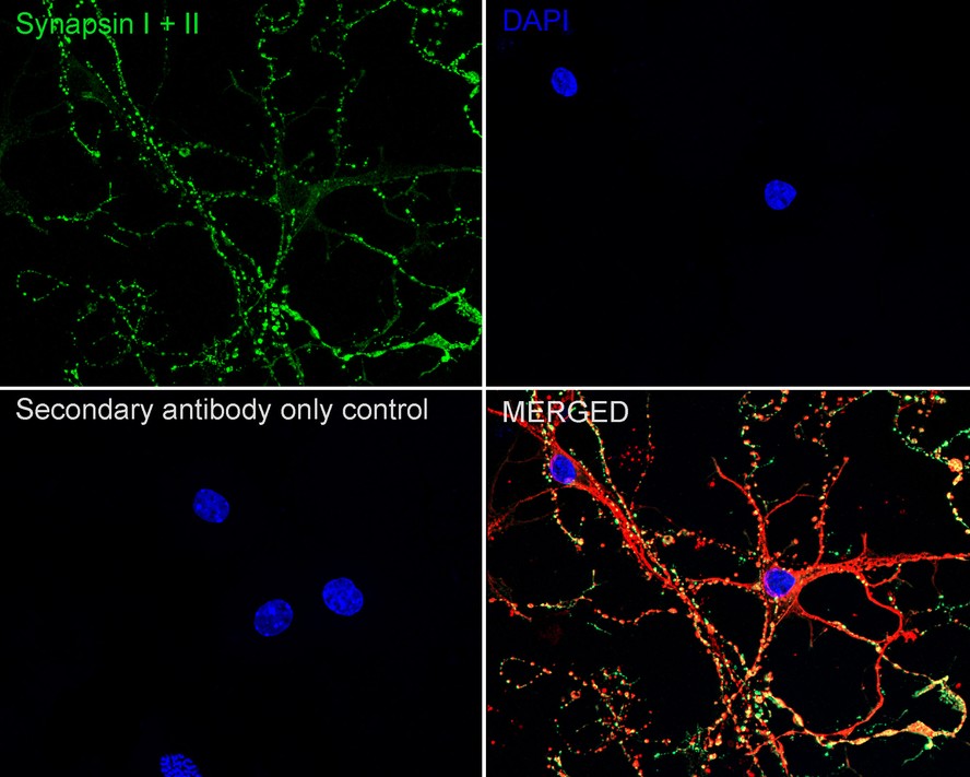
Immunocytochemistry analysis of mouse primary neuronal cells labeling Synapsin I + II with Rabbit anti-Synapsin I + II antibody at 1/500 dilution. Cells were fixed in 4% paraformaldehyde for 15 minutes at room temperature, permeabilized with 0.1% Triton X-100 in PBS for 15 minutes at room temperature, then blocked with 1% BSA in 10% negative goat serum for 1 hour at room temperature. Cells were then incubated with Rabbit anti-Synapsin I + II antibody at 1/500 dilution in 1% BSA in PBST overnight at 4 ℃. Goat Anti-Rabbit IgG H&L (488) was used as the secondary antibody at 1/1,000 dilution. PBS instead of the primary antibody was used as the secondary antibody only control. Nuclear DNA was labelled in blue with DAPI. Beta tubulin ( red) was stained at 1/100 dilution overnight at +4℃. Goat Anti-Mouse IgG H&L (594) was used as the secondary antibody at 1/1,000 dilution.IF-F
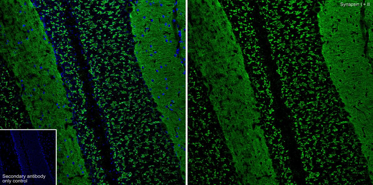
Species: Mouse, Site: Cerebellum, Sample: Frozen section, Antibody concentration: 1:500, Antigen retrieval: The section was pre-treated using heat mediated antigen retrieval with sodium citrate buffer (pH 6.0) for about 2 minutes in microwave oven.IP
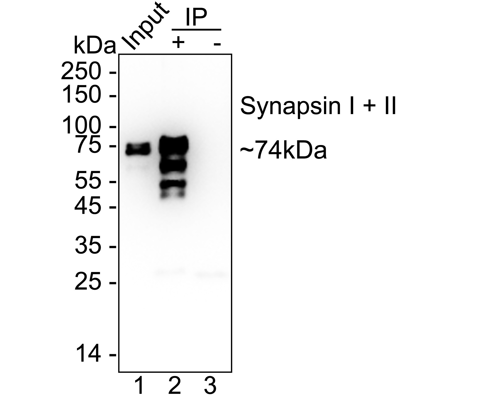
Synapsin I + II was immunoprecipitated from 0.2 mg mouse brain tissue lysate with Rabbit anti-Synapsin I + II antibody at 2 µg/10 µl beads. Western blot was performed from the immunoprecipitate using Rabbit anti-Synapsin I + II antibody at 1/1,000 dilution. Mouse Anti-Rabbit IgG kappa light chain secondary antibody at 1/5,000 dilution was used for 1 hour at room temperature. Lane 1: mouse brain tissue lysate (input), Lane 2: Rabbit anti-Synapsin I + II antibody IP in mouse brain tissue lysate, Lane 3: Rabbit IgG instead of Rabbit anti-Synapsin I + II antibody in mouse brain tissue lysate. Blocking/Dilution buffer: 5% NFDM/TBST Exposure time: 3 seconds.| Product Name | Synapsin I + II Recombinant Rabbit Monoclonal Antibody |
|---|---|
| Antibody Type | Primary Antibodies |
| Immunogen | Recombinant protein within human Synapsin I aa 1-705. |
| Clonality | monoclonal |
|---|---|
| Isotype | IgG |
| Host Species | Rabbit |
| Tested Applications | ICC/IFIF-FIHCIPWB |
| WB:1:5000 IHC:1:200-1:1000 ICC/IF:1:500 IF-F:1:500 IP:1-2μg/sample |
|
| Species Reactivity | HumanMouseRat |
| Concentration | 1mg/ml |
| Purification | Protein A |
| Gene Symbol | SYN1,SYN2 |
|---|---|
| Gene Synonyms | SYNI EPILX MRX50 SYN1a SYN1b EPILX1 |
| Gene Full Name | synapsin I,synapsin II |
| Gene Summary | This gene is a member of the synapsin gene family. Synapsins encode neuronal phosphoproteins which associate with the cytoplasmic surface of synaptic vesicles. Family members are characterized by common protein domains, and they are implicated in synaptogenesis and the modulation of neurotransmitter release, suggesting a potential role in several neuropsychiatric diseases. This member of the synapsin family plays a role in regulation of axonogenesis and synaptogenesis. The protein encoded serves as a substrate for several different protein kinases and phosphorylation may function in the regulation of this protein in the nerve terminal. Mutations in this gene may be associated with X-linked disorders with primary neuronal degeneration such as Rett syndrome. Alternatively spliced transcript variants encoding different isoforms have been identified. [provided by RefSeq, Jul 2008] |
| Molecular Weight(MW) | 74kDa(Observed band size:50-74kDa) |
| Cellular Localization | Synapse, Golgi apparatus, Presynapse, Cytoplasmic vesicle, secretory vesicle, synaptic vesicle. |
WB
Western blot analysis of Synapsin I + II on different lysates with Rabbit anti-Synapsin I + II antibody at 1/5,000 dilution. Lane 1: Mouse brain tissue lysate, Lane 2: Mouse lung tissue lysate (negative), Lane 3: Rat brain tissue lysate, Lane 4: Rat lung tissue lysate (negative), Lysates/proteins at 20 µg/Lane. Exposure time: 6 seconds; 4-20% SDS-PAGE gel. Proteins were transferred to a PVDF membrane and blocked with 5% NFDM/TBST for 1 hour at room temperature. The primary antibody at 1/5,000 dilution was used in 5% NFDM/TBST at 4℃ overnight. Goat Anti-Rabbit IgG - HRP Secondary Antibody at 1/50,000 dilution was used for 1 hour at room temperature.
IHC
Immunohistochemical analysis of paraffin-embedded mouse retina tissue with Rabbit anti-Synapsin I + II antibody at 1/200 dilution. The section was pre-treated using heat mediated antigen retrieval with Tris-EDTA buffer (pH 9.0) for 20 minutes. The tissues were blocked in 1% BSA for 20 minutes at room temperature, washed with ddH2O and PBS, and then probed with the primary antibody at 1/200 dilution for 1 hour at room temperature. The detection was performed using an HRP conjugated compact polymer system. DAB was used as the chromogen. Tissues were counterstained with hematoxylin and mounted with DPX.
ICC/IF
Immunocytochemistry analysis of mouse primary neuronal cells labeling Synapsin I + II with Rabbit anti-Synapsin I + II antibody at 1/500 dilution. Cells were fixed in 4% paraformaldehyde for 15 minutes at room temperature, permeabilized with 0.1% Triton X-100 in PBS for 15 minutes at room temperature, then blocked with 1% BSA in 10% negative goat serum for 1 hour at room temperature. Cells were then incubated with Rabbit anti-Synapsin I + II antibody at 1/500 dilution in 1% BSA in PBST overnight at 4 ℃. Goat Anti-Rabbit IgG H&L (488) was used as the secondary antibody at 1/1,000 dilution. PBS instead of the primary antibody was used as the secondary antibody only control. Nuclear DNA was labelled in blue with DAPI. Beta tubulin ( red) was stained at 1/100 dilution overnight at +4℃. Goat Anti-Mouse IgG H&L (594) was used as the secondary antibody at 1/1,000 dilution.
IF-F
Species: Mouse, Site: Cerebellum, Sample: Frozen section, Antibody concentration: 1:500, Antigen retrieval: The section was pre-treated using heat mediated antigen retrieval with sodium citrate buffer (pH 6.0) for about 2 minutes in microwave oven.
IP
Synapsin I + II was immunoprecipitated from 0.2 mg mouse brain tissue lysate with Rabbit anti-Synapsin I + II antibody at 2 µg/10 µl beads. Western blot was performed from the immunoprecipitate using Rabbit anti-Synapsin I + II antibody at 1/1,000 dilution. Mouse Anti-Rabbit IgG kappa light chain secondary antibody at 1/5,000 dilution was used for 1 hour at room temperature. Lane 1: mouse brain tissue lysate (input), Lane 2: Rabbit anti-Synapsin I + II antibody IP in mouse brain tissue lysate, Lane 3: Rabbit IgG instead of Rabbit anti-Synapsin I + II antibody in mouse brain tissue lysate. Blocking/Dilution buffer: 5% NFDM/TBST Exposure time: 3 seconds.| Application Notes | WB:1:5000 IHC:1:200-1:1000 ICC/IF:1:500 IF-F:1:500 IP:1-2μg/sample |
|---|
| Form | Liquid |
|---|---|
| Storage Instructions | Store at +4℃ after thawing. Aliquot store at -20℃. Avoid repeated freeze / thaw cycles. |
| Storage Buffer | 1*TBS (pH7.4), 0.05% BSA, 40% Glycerol. Preservative: 0.05% Sodium Azide. |


 Data sheet for OM644145