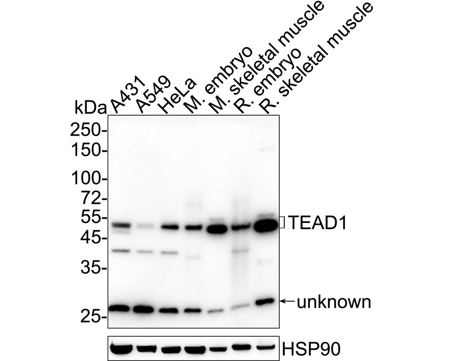
WB
Western blot analysis of TEAD1 on different lysates with Rabbit anti-TEAD1 antibody at 1/1,000 dilution. Lane 1: A431 cell lysate, Lane 2: A549 cell lysate, Lane 3: HeLa cell lysate, Lane 4: Mouse embryo tissue lysate, Lane 5: Mouse skeletal muscle tissue lysate, Lane 6: Rat embryo tissue lysate, Lane 7: Rat skeletal muscle tissue lysate, Lysates/proteins at 20 µg/Lane. Exposure time: 2 minutes 7 seconds; 4-20% SDS-PAGE gel. Proteins were transferred to a PVDF membrane and blocked with 5% NFDM/TBST for 1 hour at room temperature. The primary antibody at 1/1,000 dilution was used in 5% NFDM/TBST at 4℃ overnight. Goat Anti-Rabbit IgG - HRP Secondary Antibody at 1/50,000 dilution was used for 1 hour at room temperature.IHC
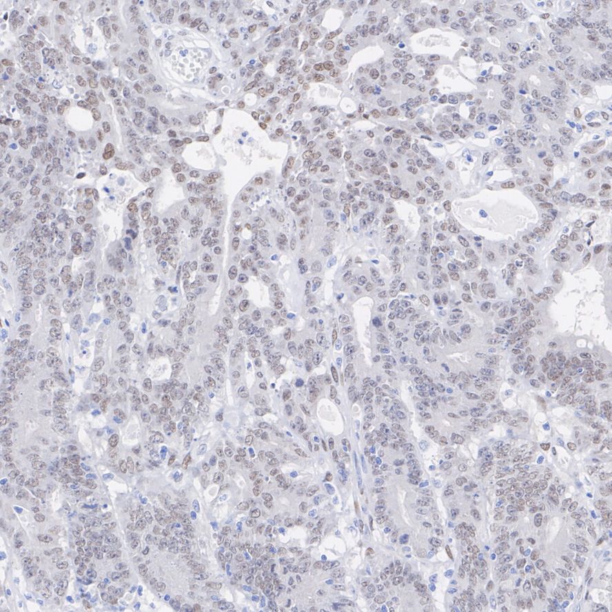
Immunohistochemical analysis of paraffin-embedded human colon cancer tissue with Rabbit anti-TEAD1 antibody at 1/200 dilution. The section was pre-treated using heat mediated antigen retrieval with sodium citrate buffer (pH 6.0) for 2 minutes. The tissues were blocked in 1% BSA for 20 minutes at room temperature, washed with ddH2O and PBS, and then probed with the primary antibody at 1/200 dilution for 1 hour at room temperature. The detection was performed using an HRP conjugated compact polymer system. DAB was used as the chromogen. Tissues were counterstained with hematoxylin and mounted with DPX.ICC/IF
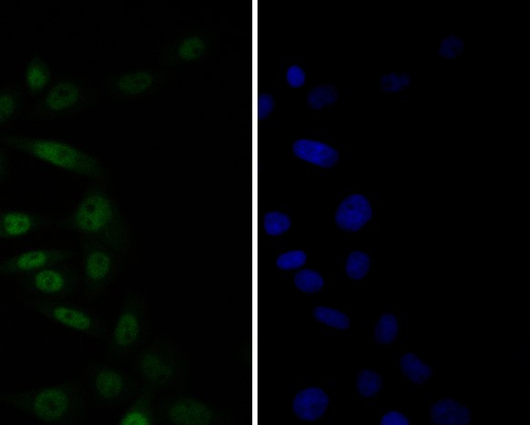
Immunocytochemistry analysis of SiHa cells labeling TEAD1 with Rabbit anti-TEAD1 antibody at 1/50 dilution. Cells were fixed in 4% paraformaldehyde for 10 minutes at 37 ℃, permeabilized with 0.05% Triton X-100 in PBS for 20 minutes, and then blocked with 2% negative goat serum for 30 minutes at room temperature. Cells were then incubated with Rabbit anti-TEAD1 antibody at 1/50 dilution in 2% negative goat serum overnight at 4 ℃.Alexa Fluor®488 Goat anti-Rabbit IgG was used as the secondary antibody at 1/1,000 dilution. Nuclear DNA was labelled in blue with DAPI.| Product Name | TEAD1 Recombinant Rabbit Monoclonal Antibody |
|---|---|
| Antibody Type | Primary Antibodies |
| Immunogen | Synthetic peptide within Human TEAD1 aa 1-50 / 426. |
| Clonality | monoclonal |
|---|---|
| Isotype | IgG |
| Host Species | Rabbit |
| Tested Applications | ICC/IFIHCWB |
| WB:1:1000 IHC:1:50-1:200 ICC/IF:1:50-1:100 |
|
| Species Reactivity | HumanMouseRat |
| Concentration | 1mg/ml |
| Purification | Protein A |
| Gene Symbol | TEAD1 |
|---|---|
| Gene Synonyms | AA REF1 TCF13 TEF-1 NTEF-1 TCF-13 TEAD-1 |
| Gene Full Name | TEA domain transcription factor 1 |
| Gene Summary | This gene encodes a ubiquitous transcriptional enhancer factor that is a member of the TEA/ATTS domain family. This protein directs the transactivation of a wide variety of genes and, in placental cells, also acts as a transcriptional repressor. Mutations in this gene cause Sveinsson's chorioretinal atrophy. Additional transcript variants have been described but their full-length natures have not been experimentally verified. [provided by RefSeq, May 2010] |
| Molecular Weight(MW) | 48kDa(Observed band size:48/50kDa) |
| Cellular Localization | Nucleus. |
WB
Western blot analysis of TEAD1 on different lysates with Rabbit anti-TEAD1 antibody at 1/1,000 dilution. Lane 1: A431 cell lysate, Lane 2: A549 cell lysate, Lane 3: HeLa cell lysate, Lane 4: Mouse embryo tissue lysate, Lane 5: Mouse skeletal muscle tissue lysate, Lane 6: Rat embryo tissue lysate, Lane 7: Rat skeletal muscle tissue lysate, Lysates/proteins at 20 µg/Lane. Exposure time: 2 minutes 7 seconds; 4-20% SDS-PAGE gel. Proteins were transferred to a PVDF membrane and blocked with 5% NFDM/TBST for 1 hour at room temperature. The primary antibody at 1/1,000 dilution was used in 5% NFDM/TBST at 4℃ overnight. Goat Anti-Rabbit IgG - HRP Secondary Antibody at 1/50,000 dilution was used for 1 hour at room temperature.
IHC
Immunohistochemical analysis of paraffin-embedded human colon cancer tissue with Rabbit anti-TEAD1 antibody at 1/200 dilution. The section was pre-treated using heat mediated antigen retrieval with sodium citrate buffer (pH 6.0) for 2 minutes. The tissues were blocked in 1% BSA for 20 minutes at room temperature, washed with ddH2O and PBS, and then probed with the primary antibody at 1/200 dilution for 1 hour at room temperature. The detection was performed using an HRP conjugated compact polymer system. DAB was used as the chromogen. Tissues were counterstained with hematoxylin and mounted with DPX.
ICC/IF
Immunocytochemistry analysis of SiHa cells labeling TEAD1 with Rabbit anti-TEAD1 antibody at 1/50 dilution. Cells were fixed in 4% paraformaldehyde for 10 minutes at 37 ℃, permeabilized with 0.05% Triton X-100 in PBS for 20 minutes, and then blocked with 2% negative goat serum for 30 minutes at room temperature. Cells were then incubated with Rabbit anti-TEAD1 antibody at 1/50 dilution in 2% negative goat serum overnight at 4 ℃.Alexa Fluor®488 Goat anti-Rabbit IgG was used as the secondary antibody at 1/1,000 dilution. Nuclear DNA was labelled in blue with DAPI.| Application Notes | WB:1:1000 IHC:1:50-1:200 ICC/IF:1:50-1:100 |
|---|
| Form | Liquid |
|---|---|
| Storage Instructions | Store at +4℃ after thawing. Aliquot store at -20℃. Avoid repeated freeze / thaw cycles. |
| Storage Buffer | 1*TBS (pH7.4), 0.05% BSA, 40% Glycerol. Preservative: 0.05% Sodium Azide. |


 Data sheet for OM644155