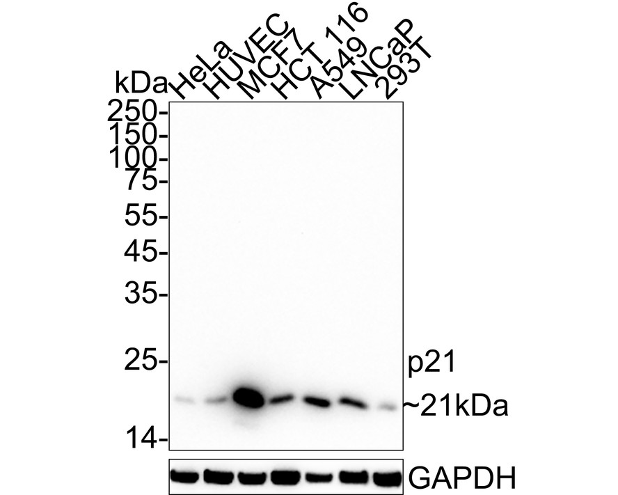
WB
Western blot analysis of p21 on different lysates with Rabbit anti-p21 antibody at 1/1,000 dilution. Lane 1: HeLa cell lysate, Lane 2: HUVEC cell lysate, Lane 3: MCF7 cell lysate, Lane 4: HCT 116 cell lysate, Lane 5: A549 cell lysate, Lane 6: LNCaP cell lysate, Lane 7: 293T cell lysate, Lysates/proteins at 20 µg/Lane. Exposure time: 3 minutes; 4-20% SDS-PAGE gel. Proteins were transferred to a PVDF membrane and blocked with 5% NFDM/TBST for 1 hour at room temperature. The primary antibody at 1/1,000 dilution was used in 5% NFDM/TBST at 4℃ overnight. Goat Anti-Rabbit IgG - HRP Secondary Antibody at 1/50,000 dilution was used for 1 hour at room temperature.IHC
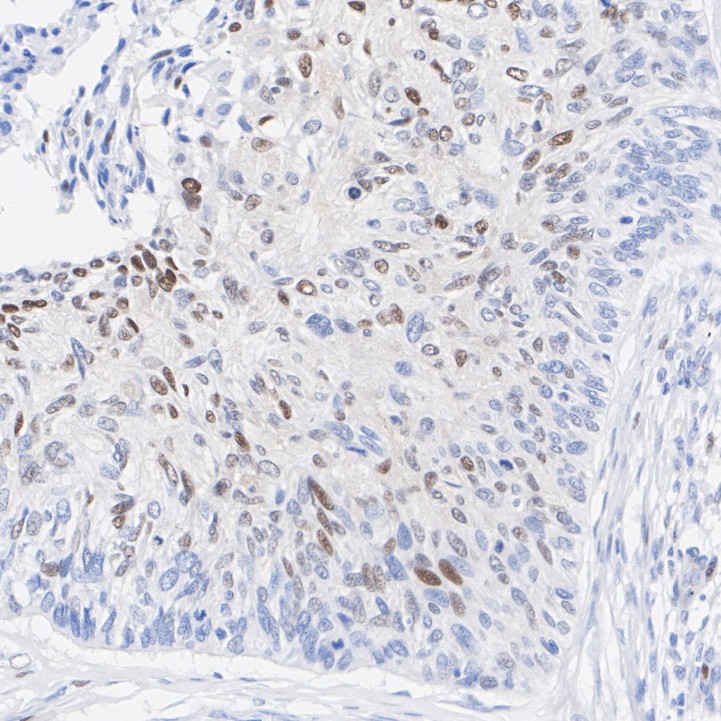
Immunohistochemical analysis of paraffin-embedded human cervical cancer tissue with Rabbit anti-p21 antibody at 1/50 dilution. The section was pre-treated using heat mediated antigen retrieval with sodium citrate buffer (pH 6.0) for 2 minutes. The tissues were blocked in 1% BSA for 20 minutes at room temperature, washed with ddH2O and PBS, and then probed with the primary antibody at 1/50 dilution for 1 hour at room temperature. The detection was performed using an HRP conjugated compact polymer system. DAB was used as the chromogen. Tissues were counterstained with hematoxylin and mounted with DPX.ICC/IF
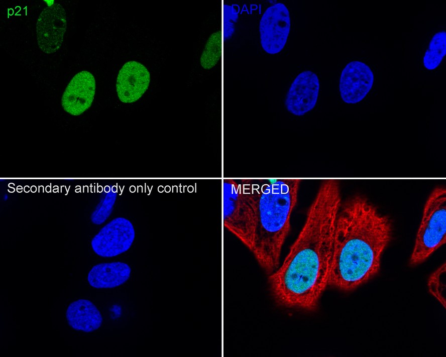
Immunocytochemistry analysis of MCF7 cells labeling p21 with Rabbit anti-p21 antibody at 1/100 dilution. Cells were fixed in 4% paraformaldehyde for 20 minutes at room temperature, permeabilized with 0.1% Triton X-100 in PBS for 5 minutes at room temperature, then blocked with 1% BSA in 10% negative goat serum for 1 hour at room temperature. Cells were then incubated with Rabbit anti-p21 antibody at 1/100 dilution in 1% BSA in PBST overnight at 4 ℃. Goat Anti-Rabbit IgG H&L (488) was used as the secondary antibody at 1/1,000 dilution. PBS instead of the primary antibody was used as the secondary antibody only control. Nuclear DNA was labelled in blue with DAPI. Beta tubulin (red) was stained at 1/100 dilution overnight at +4℃. Goat Anti-Mouse IgG H&L (594) was used as the secondary antibody at 1/1,000 dilution.IP
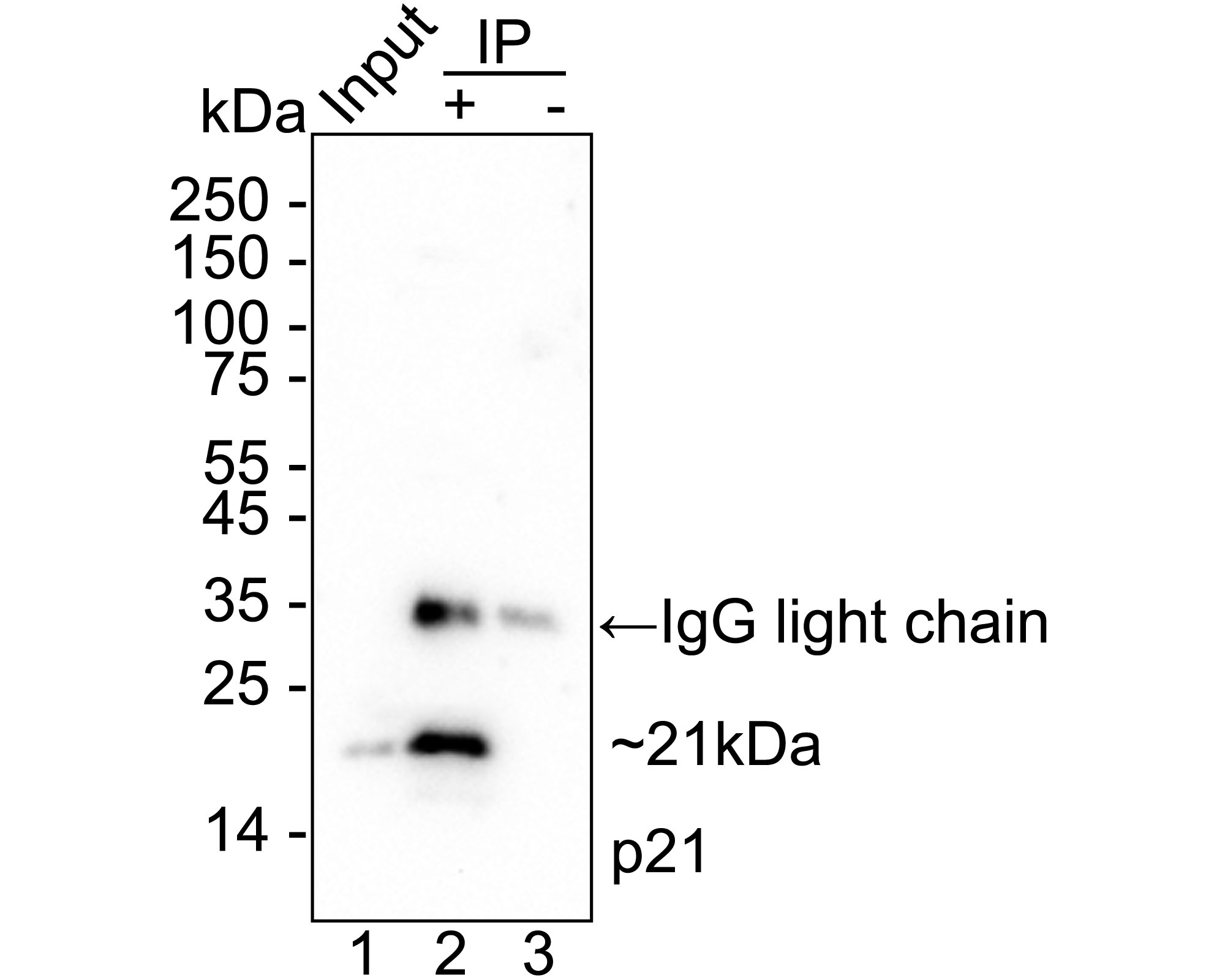
p21 was immunoprecipitated from 0.2 mg MCF7 cell lysate with Rabbit anti-p21 antibody at 2 µg/25 µl agarose. Western blot was performed from the immunoprecipitate using Rabbit anti-p21 antibody at 1/1,000 dilution. Anti-Rabbit IgG for IP Nano-secondary antibody at 1/5,000 dilution was used for 1 hour at room temperature. Lane 1: MCF7 cell lysate (input), Lane 2: Rabbit anti-p21 antibody IP in MCF7 cell lysate, Lane 3: Rabbit IgG instead of Rabbit anti-p21 antibody in MCF7 cell lysate, Blocking/Dilution buffer: 5% NFDM/TBST, Exposure time: 1 minute 31 seconds.| Product Name | p21 Recombinant Rabbit Monoclonal Antibody |
|---|---|
| Antibody Type | Primary Antibodies |
| Immunogen | Recombinant protein within |
| Clonality | monoclonal |
|---|---|
| Isotype | IgG |
| Host Species | Rabbit |
| Tested Applications | ICC/IFIHCIPWB |
| WB:1:1000 IHC:1:50 ICC/IF:1:100 IP:1-2μg/sample |
|
| Species Reactivity | Human |
| Concentration | 1mg/ml |
| Purification | Protein A |
| Gene Symbol | CDKN1A |
|---|---|
| Gene Synonyms | P21 CIP1 SDI1 WAF1 CAP20 CDKN1 MDA-6 p21CIP1 |
| Gene Full Name | cyclin dependent kinase inhibitor 1A |
| Gene Summary | This gene encodes a potent cyclin-dependent kinase inhibitor. The encoded protein binds to and inhibits the activity of cyclin-cyclin-dependent kinase2 or -cyclin-dependent kinase4 complexes, and thus functions as a regulator of cell cycle progression at G1. The expression of this gene is tightly controlled by the tumor suppressor protein p53, through which this protein mediates the p53-dependent cell cycle G1 phase arrest in response to a variety of stress stimuli. This protein can interact with proliferating cell nuclear antigen, a DNA polymerase accessory factor, and plays a regulatory role in S phase DNA replication and DNA damage repair. This protein was reported to be specifically cleaved by CASP3-like caspases, which thus leads to a dramatic activation of cyclin-dependent kinase2, and may be instrumental in the execution of apoptosis following caspase activation. Mice that lack this gene have the ability to regenerate damaged or missing tissue. Multiple alternatively spliced variants have been found for this gene. [provided by RefSeq, Sep 2015] |
| Molecular Weight(MW) | 18kDa(Observed band size: 21kDa) |
| Cellular Localization | Cytoplasm, Nucleus. |
WB
Western blot analysis of p21 on different lysates with Rabbit anti-p21 antibody at 1/1,000 dilution. Lane 1: HeLa cell lysate, Lane 2: HUVEC cell lysate, Lane 3: MCF7 cell lysate, Lane 4: HCT 116 cell lysate, Lane 5: A549 cell lysate, Lane 6: LNCaP cell lysate, Lane 7: 293T cell lysate, Lysates/proteins at 20 µg/Lane. Exposure time: 3 minutes; 4-20% SDS-PAGE gel. Proteins were transferred to a PVDF membrane and blocked with 5% NFDM/TBST for 1 hour at room temperature. The primary antibody at 1/1,000 dilution was used in 5% NFDM/TBST at 4℃ overnight. Goat Anti-Rabbit IgG - HRP Secondary Antibody at 1/50,000 dilution was used for 1 hour at room temperature.
IHC
Immunohistochemical analysis of paraffin-embedded human cervical cancer tissue with Rabbit anti-p21 antibody at 1/50 dilution. The section was pre-treated using heat mediated antigen retrieval with sodium citrate buffer (pH 6.0) for 2 minutes. The tissues were blocked in 1% BSA for 20 minutes at room temperature, washed with ddH2O and PBS, and then probed with the primary antibody at 1/50 dilution for 1 hour at room temperature. The detection was performed using an HRP conjugated compact polymer system. DAB was used as the chromogen. Tissues were counterstained with hematoxylin and mounted with DPX.
ICC/IF
Immunocytochemistry analysis of MCF7 cells labeling p21 with Rabbit anti-p21 antibody at 1/100 dilution. Cells were fixed in 4% paraformaldehyde for 20 minutes at room temperature, permeabilized with 0.1% Triton X-100 in PBS for 5 minutes at room temperature, then blocked with 1% BSA in 10% negative goat serum for 1 hour at room temperature. Cells were then incubated with Rabbit anti-p21 antibody at 1/100 dilution in 1% BSA in PBST overnight at 4 ℃. Goat Anti-Rabbit IgG H&L (488) was used as the secondary antibody at 1/1,000 dilution. PBS instead of the primary antibody was used as the secondary antibody only control. Nuclear DNA was labelled in blue with DAPI. Beta tubulin (red) was stained at 1/100 dilution overnight at +4℃. Goat Anti-Mouse IgG H&L (594) was used as the secondary antibody at 1/1,000 dilution.
IP
p21 was immunoprecipitated from 0.2 mg MCF7 cell lysate with Rabbit anti-p21 antibody at 2 µg/25 µl agarose. Western blot was performed from the immunoprecipitate using Rabbit anti-p21 antibody at 1/1,000 dilution. Anti-Rabbit IgG for IP Nano-secondary antibody at 1/5,000 dilution was used for 1 hour at room temperature. Lane 1: MCF7 cell lysate (input), Lane 2: Rabbit anti-p21 antibody IP in MCF7 cell lysate, Lane 3: Rabbit IgG instead of Rabbit anti-p21 antibody in MCF7 cell lysate, Blocking/Dilution buffer: 5% NFDM/TBST, Exposure time: 1 minute 31 seconds.| Application Notes | WB:1:1000 IHC:1:50 ICC/IF:1:100 IP:1-2μg/sample |
|---|
| Form | Liquid |
|---|---|
| Storage Instructions | Store at +4℃ after thawing. Aliquot store at -20℃. Avoid repeated freeze / thaw cycles. |
| Storage Buffer | 1*TBS (pH7.4), 0.05% BSA, 40% Glycerol. Preservative: 0.05% Sodium Azide. |


 Data sheet for OM644192