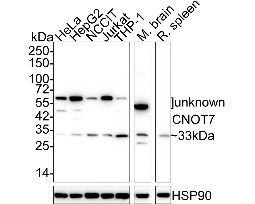

WB
Western blot analysis of CNOT7 on different lysates with Rabbit anti-CNOT7 antibody at 1/2,000 dilution. Lane 1: HeLa cell lysate (20 µg/Lane), Lane 2: HepG2 cell lysate (20 µg/Lane), Lane 3: NCCIT cell lysate (20 µg/Lane), Lane 4: Jurkat cell lysate (20 µg/Lane), Lane 5: THP-1 cell lysate (20 µg/Lane), Lane 6: Mouse brain tissue lysate (40 µg/Lane), Lane 7: Rat spleen tissue lysate (40 µg/Lane), Exposure time: 3 minutes 20 seconds; 4-20% SDS-PAGE gel. Proteins were transferred to a PVDF membrane and blocked with 5% NFDM/TBST for 1 hour at room temperature. The primary antibody at 1/2,000 dilution was used in 5% NFDM/TBST at 4℃ overnight. Goat Anti-Rabbit IgG - HRP Secondary Antibody at 1/50,000 dilution was used for 1 hour at room temperature.ICC/IF
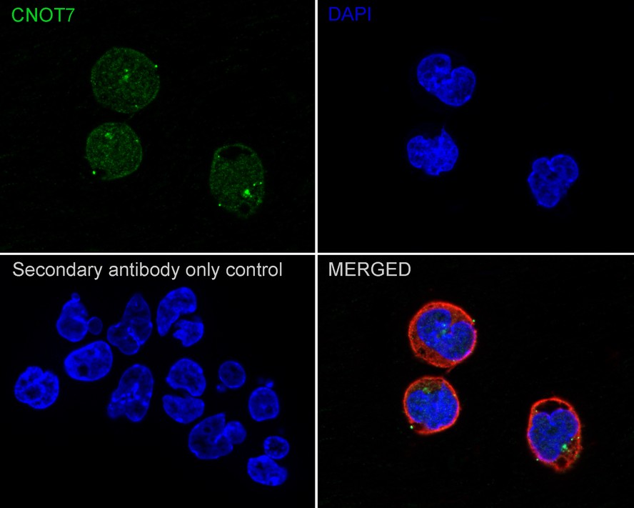
Immunocytochemistry analysis of F9 cells labeling CNOT7 with Rabbit anti-CNOT7 antibody at 1/200 dilution. Cells were fixed in 4% paraformaldehyde for 20 minutes at room temperature, permeabilized with 0.1% Triton X-100 in PBS for 5 minutes at room temperature, then blocked with 1% BSA in 10% negative goat serum for 1 hour at room temperature. Cells were then incubated with Rabbit anti-CNOT7 antibody at 1/200 dilution in 1% BSA in PBST overnight at 4 ℃. Goat Anti-Rabbit IgG H&L (488) was used as the secondary antibody at 1/1,000 dilution. PBS instead of the primary antibody was used as the secondary antibody only control. Nuclear DNA was labelled in blue with DAPI. Beta tubulin (red) was stained at 1/100 dilution overnight at +4℃. Goat Anti-Mouse IgG H&L (594) was used as the secondary antibody at 1/1,000 dilution.FC
Flow cytometric analysis of F9 cells labeling CNOT7 Cells were fixed and permeabilized. Then stained with the primary antibody (1μg/mL) (red) compared with Rabbit IgG Isotype Control (green). After incubation of the primary antibody at +4℃ for an hour, the cells were stained with a iFluor™ 488 conjugate-Goat anti-Rabbit IgG Secondary antibody at 1/1,000 dilution for 30 minutes at +4℃. Unlabelled sample was used as a control (cells without incubation with primary antibody; black).| Product Name | CNOT7 Recombinant Rabbit Monoclonal Antibody |
|---|---|
| Antibody Type | Primary Antibodies |
| Immunogen | Recombinant protein within human CNOT7 aa 1-285 / 285. |
| Clonality | monoclonal |
|---|---|
| Isotype | IgG |
| Host Species | Rabbit |
| Tested Applications | FCICC/IFWB |
| WB:1:2000 ICC/IF:1:100 FC:1:1000 |
|
| Species Reactivity | HumanMouseRat |
| Concentration | 1mg/ml |
| Purification | Protein A |
| Gene Symbol | CNOT7 |
|---|---|
| Gene Synonyms | CAF1 CAF-1 Caf1a hCAF-1 |
| Gene Full Name | CCR4-NOT transcription complex subunit 7 |
| Gene Summary | The protein encoded by this gene binds to an anti-proliferative protein, B-cell translocation protein 1, which negatively regulates cell proliferation. Binding of the two proteins, which is driven by phosphorylation of the anti-proliferative protein, causes signaling events in cell division that lead to changes in cell proliferation associated with cell-cell contact. The encoded protein downregulates the innate immune response and therefore provides a therapeutic target for enhancing its antimicrobial activity against foreign agents. Alternative splicing of this gene results in multiple transcript variants. Related pseudogenes have been identified on chromosomes 1 and X. [provided by RefSeq, Apr 2016] |
| Molecular Weight(MW) | 33kDa |
| Cellular Localization | Nucleus, Cytoplasm, P-body, Cytoplasmic ribonucleoprotein granule. |
WB
Western blot analysis of CNOT7 on different lysates with Rabbit anti-CNOT7 antibody at 1/2,000 dilution. Lane 1: HeLa cell lysate (20 µg/Lane), Lane 2: HepG2 cell lysate (20 µg/Lane), Lane 3: NCCIT cell lysate (20 µg/Lane), Lane 4: Jurkat cell lysate (20 µg/Lane), Lane 5: THP-1 cell lysate (20 µg/Lane), Lane 6: Mouse brain tissue lysate (40 µg/Lane), Lane 7: Rat spleen tissue lysate (40 µg/Lane), Exposure time: 3 minutes 20 seconds; 4-20% SDS-PAGE gel. Proteins were transferred to a PVDF membrane and blocked with 5% NFDM/TBST for 1 hour at room temperature. The primary antibody at 1/2,000 dilution was used in 5% NFDM/TBST at 4℃ overnight. Goat Anti-Rabbit IgG - HRP Secondary Antibody at 1/50,000 dilution was used for 1 hour at room temperature.
ICC/IF
Immunocytochemistry analysis of F9 cells labeling CNOT7 with Rabbit anti-CNOT7 antibody at 1/200 dilution. Cells were fixed in 4% paraformaldehyde for 20 minutes at room temperature, permeabilized with 0.1% Triton X-100 in PBS for 5 minutes at room temperature, then blocked with 1% BSA in 10% negative goat serum for 1 hour at room temperature. Cells were then incubated with Rabbit anti-CNOT7 antibody at 1/200 dilution in 1% BSA in PBST overnight at 4 ℃. Goat Anti-Rabbit IgG H&L (488) was used as the secondary antibody at 1/1,000 dilution. PBS instead of the primary antibody was used as the secondary antibody only control. Nuclear DNA was labelled in blue with DAPI. Beta tubulin (red) was stained at 1/100 dilution overnight at +4℃. Goat Anti-Mouse IgG H&L (594) was used as the secondary antibody at 1/1,000 dilution.
FC
Flow cytometric analysis of F9 cells labeling CNOT7 Cells were fixed and permeabilized. Then stained with the primary antibody (1μg/mL) (red) compared with Rabbit IgG Isotype Control (green). After incubation of the primary antibody at +4℃ for an hour, the cells were stained with a iFluor™ 488 conjugate-Goat anti-Rabbit IgG Secondary antibody at 1/1,000 dilution for 30 minutes at +4℃. Unlabelled sample was used as a control (cells without incubation with primary antibody; black).| Application Notes | WB:1:2000 ICC/IF:1:100 FC:1:1000 |
|---|
| Form | Liquid |
|---|---|
| Storage Instructions | Store at +4℃ after thawing. Aliquot store at -20℃. Avoid repeated freeze / thaw cycles. |
| Storage Buffer | PBS (pH7.4), 0.1% BSA, 40% Glycerol. Preservative: 0.05% Sodium Azide. |


 Data sheet for OM644287