WB
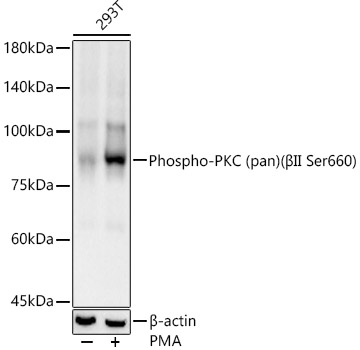
Western blot analysis of lysates from 293T cells, using Phospho-PKC (pan) (βII Ser660) Rabbit mAb at 1:1000 dilution.293T cells were treated by PMA/TPA (200 nM) at 37℃ for 30 minutes after serum-starvation overnight.Secondary antibody: HRP-conjugated Goat anti-Rabbit IgG (H+L) at 1:10000 dilution.Lysates/proteins: 25μg per lane.Blocking buffer: 3% nonfat dry milk in TBST.Detection: ECL Basic Kit.Exposure time: 30s.WB
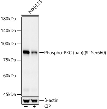
Western blot analysis of lysates from NIH/3T3 cells, using Phospho-PKC (pan) (βII Ser660) Rabbit mAb at 1:1000 dilution.NIH/3T3 cells were treated by CIP(20uL/400ul) at 37℃ for 1 hour.Secondary antibody: HRP-conjugated Goat anti-Rabbit IgG (H+L) at 1:10000 dilution.Lysates/proteins: 25μg per lane.Blocking buffer: 3% nonfat dry milk in TBST.Detection: ECL Basic Kit.Exposure time: 30s.IHC
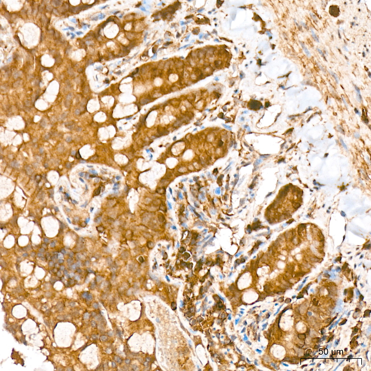
Immunohistochemistry analysis of paraffin embedded Rat colon tissue using Phospho PKC (pan) (βII Ser660) Rabbit mAb at a dilution of 1:400 (40x lens). High pressure antigen retrieval was performed with 0.01 M citrate buffer (pH 6.0) prior to IHC staining.IHC
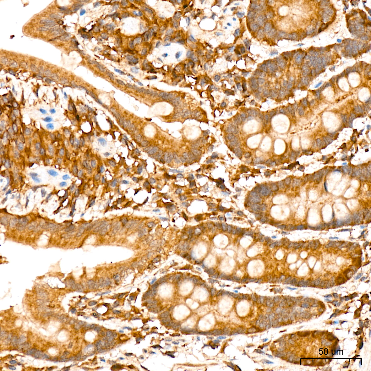
Immunohistochemistry analysis of paraffin embedded Human colon tissue using Phospho-PKC (pan) (βII Ser660) Rabbit mAb at a dilution of 1:400 (40x lens). High pressure antigen retrieval was performed with 0.01 M citrate buffer (pH 6.0) prior to IHC staining.| Product Name | Phospho-PKC (pan) (βII Ser660) Rabbit mAb |
|---|---|
| Antibody Type | Primary Antibodies |
| Immunogen | A synthetic phosphorylated peptide around S660 of human PRKCA(NP_002728.2). |
| Modification | p-βII Ser660 |
| Clonality | monoclonal |
|---|---|
| Isotype | IgG |
| Host Species | Rabbit |
| Tested Applications | IHCWB |
| WB:1:500-1:1000 IHC:1:100-1:500 |
|
| Species Reactivity | HumanMouseRat |
| Concentration | 1mg/ml |
| Purification | Affinity purified |
| Gene Symbol | PRKCA |
|---|---|
| Gene Synonyms | AAG6 PKCA PRKACA PKCI+/- PKCalpha PKC-alpha |
| Gene Full Name | protein kinase C alpha |
| Gene Summary | Protein kinase C (PKC) is a family of serine- and threonine-specific protein kinases that can be activated by calcium and the second messenger diacylglycerol. PKC family members phosphorylate a wide variety of protein targets and are known to be involved in diverse cellular signaling pathways. PKC family members also serve as major receptors for phorbol esters, a class of tumor promoters. Each member of the PKC family has a specific expression profile and is believed to play a distinct role in cells. The protein encoded by this gene is one of the PKC family members. This kinase has been reported to play roles in many different cellular processes, such as cell adhesion, cell transformation, cell cycle checkpoint, and cell volume control. Knockout studies in mice suggest that this kinase may be a fundamental regulator of cardiac contractility and Ca(2+) handling in myocytes. [provided by RefSeq, Jul 2008] |
| Molecular Weight(MW) | 85kDa |
| Cellular Localization | Cell membrane,Cytoplasm,Membrane,Mitochondrion,Nucleus. |
WB
Western blot analysis of lysates from 293T cells, using Phospho-PKC (pan) (βII Ser660) Rabbit mAb at 1:1000 dilution.293T cells were treated by PMA/TPA (200 nM) at 37℃ for 30 minutes after serum-starvation overnight.Secondary antibody: HRP-conjugated Goat anti-Rabbit IgG (H+L) at 1:10000 dilution.Lysates/proteins: 25μg per lane.Blocking buffer: 3% nonfat dry milk in TBST.Detection: ECL Basic Kit.Exposure time: 30s.
WB
Western blot analysis of lysates from NIH/3T3 cells, using Phospho-PKC (pan) (βII Ser660) Rabbit mAb at 1:1000 dilution.NIH/3T3 cells were treated by CIP(20uL/400ul) at 37℃ for 1 hour.Secondary antibody: HRP-conjugated Goat anti-Rabbit IgG (H+L) at 1:10000 dilution.Lysates/proteins: 25μg per lane.Blocking buffer: 3% nonfat dry milk in TBST.Detection: ECL Basic Kit.Exposure time: 30s.
IHC
Immunohistochemistry analysis of paraffin embedded Rat colon tissue using Phospho PKC (pan) (βII Ser660) Rabbit mAb at a dilution of 1:400 (40x lens). High pressure antigen retrieval was performed with 0.01 M citrate buffer (pH 6.0) prior to IHC staining.
IHC
Immunohistochemistry analysis of paraffin embedded Human colon tissue using Phospho-PKC (pan) (βII Ser660) Rabbit mAb at a dilution of 1:400 (40x lens). High pressure antigen retrieval was performed with 0.01 M citrate buffer (pH 6.0) prior to IHC staining.| Application Notes | WB:1:500-1:1000 IHC:1:100-1:500 |
|---|
| Form | Liquid |
|---|---|
| Storage Instructions | Store at -20℃. Avoid freeze / thaw cycles. |
| Storage Buffer | PBS with 0.05% proclin300, 0.05% BSA, 50% glycerol, pH7.3. |


 Data sheet for OM644357