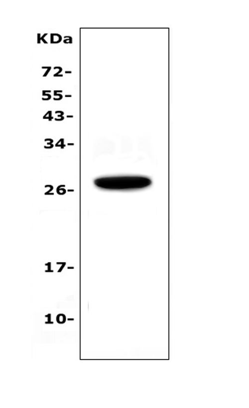
WB
Western blot analysis of IL15RA using anti-IL15RA antibody. The sample well of each lane was loaded with 30 ug of sample under reducing conditions. Lane 1: human placenta tissue lysates. After electrophoresis, proteins were transferred to a membrane. Then the membrane was incubated with rabbit anti-IL15RA antigen affinity purified polyclonal antibody at a dilution of 1:1000 and probed with a goat anti-rabbit IgG-HRP secondary antibody. The signal is developed using ECL Plus Western Blotting Substrate.IHC
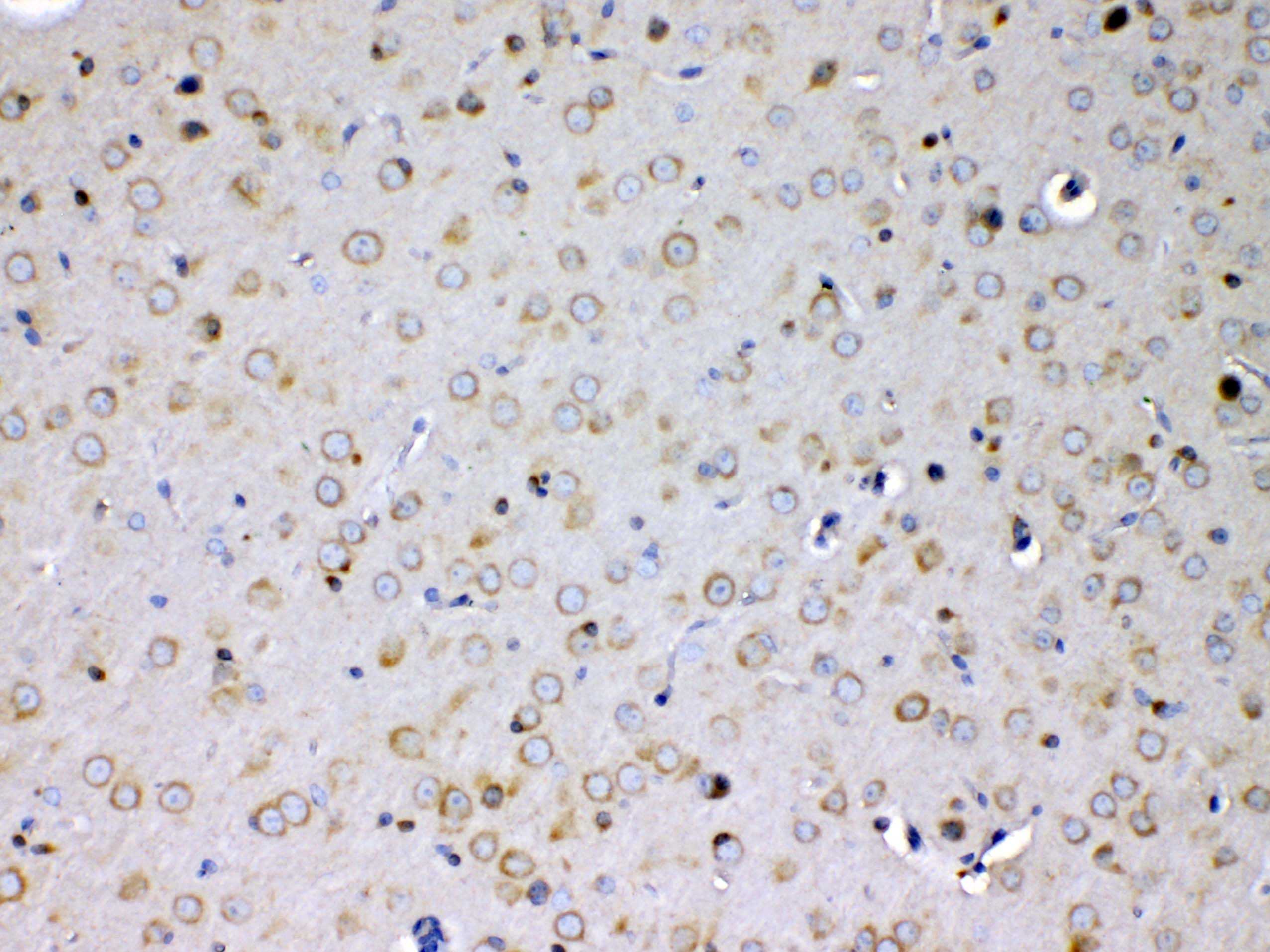
IHC analysis of IL15RA using anti-IL15RA antibody. IL15RA was detected in a paraffin-embedded section of mouse brain tissue. Biotinylated goat anti-rabbit IgG was used as secondary antibody. The tissue section was incubated with rabbit anti-IL15RA Antibody at a dilution of 1:200 and developed using Strepavidin-Biotin-Complex (SABC) with DAB as the chromogen.ICC/IF
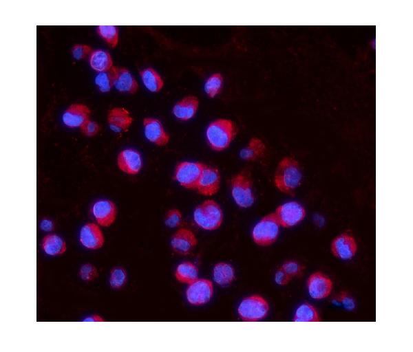
IF analysis of IL15RA using anti-IL15RA antibody. IL15RA was detected in an immunocytochemical section of K562 cells. Dylight594-conjugated Anti-rabbit IgG Secondary Antibody (red) was used as secondary antibody. The section was counterstained with DAPI (Blue).FC
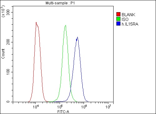
Flow Cytometry analysis of K562 cells using anti-IL15RA antibody. Overlay histogram showing K562 cells stained with anti-IL15RA antibody (Blue line). To facilitate intracellular staining, cells were fixed with 4% paraformaldehyde and permeabilized with permeabilization buffer. The cells were blocked with 10% normal goat serum. And then incubated with rabbit anti-IL15RA Antibody at 1:100 dilution for 30 min at 20°C. DyLight®488 conjugated goat anti-rabbit IgG was used as secondary antibody at 1:100 dilution for 30 minutes at 20°C. Isotype control antibody (Green line) was rabbit IgG at 1:100 dilution used under the same conditions. Unlabelled sample without incubation with primary antibody and secondary antibody (Red line) was used as a blank control.| Product Name | Rabbit polyclonal antibody to IL15RA |
|---|---|
| Antibody Type | Primary Antibodies |
| Immunogen | E.coli-derived human IL15RA recombinant protein (Position: I31-T174). Human IL15RA shares 67.9% and 68.8% amino acid (aa) sequence identity with mouse and rat IL15RA, respectively. |
| Clonality | polyclonal |
|---|---|
| Isotype | IgG |
| Host Species | Rabbit |
| Tested Applications | FCICC/IFIHCWB |
| WB:1:500-1:2000 IHC:1:50-1:400 ICC/IF:1:50-1:400 FC:1:50-1:200 |
|
| Species Reactivity | HumanMouseRat |
| Concentration | 0.5mg/ml |
| Purification | Affinity purified |
| Gene Symbol | IL15RA |
|---|---|
| Gene Synonyms | CD215 |
| Gene Full Name | interleukin 15 receptor subunit alpha |
| Gene Summary | This gene encodes a cytokine receptor that specifically binds interleukin 15 (IL15) with high affinity. The receptors of IL15 and IL2 share two subunits, IL2R beta and IL2R gamma. This forms the basis of many overlapping biological activities of IL15 and IL2. The protein encoded by this gene is structurally related to IL2R alpha, an additional IL2-specific alpha subunit necessary for high affinity IL2 binding. Unlike IL2RA, IL15RA is capable of binding IL15 with high affinity independent of other subunits, which suggests distinct roles between IL15 and IL2. This receptor is reported to enhance cell proliferation and expression of apoptosis inhibitor BCL2L1/BCL2-XL and BCL2. Multiple alternatively spliced transcript variants of this gene have been reported.[provided by RefSeq, Apr 2010] |
| Molecular Weight(MW) | 28kDa |
| Cellular Localization | Membrane. |
WB
Western blot analysis of IL15RA using anti-IL15RA antibody. The sample well of each lane was loaded with 30 ug of sample under reducing conditions. Lane 1: human placenta tissue lysates. After electrophoresis, proteins were transferred to a membrane. Then the membrane was incubated with rabbit anti-IL15RA antigen affinity purified polyclonal antibody at a dilution of 1:1000 and probed with a goat anti-rabbit IgG-HRP secondary antibody. The signal is developed using ECL Plus Western Blotting Substrate.
IHC
IHC analysis of IL15RA using anti-IL15RA antibody. IL15RA was detected in a paraffin-embedded section of mouse brain tissue. Biotinylated goat anti-rabbit IgG was used as secondary antibody. The tissue section was incubated with rabbit anti-IL15RA Antibody at a dilution of 1:200 and developed using Strepavidin-Biotin-Complex (SABC) with DAB as the chromogen.
ICC/IF
IF analysis of IL15RA using anti-IL15RA antibody. IL15RA was detected in an immunocytochemical section of K562 cells. Dylight594-conjugated Anti-rabbit IgG Secondary Antibody (red) was used as secondary antibody. The section was counterstained with DAPI (Blue).
FC
Flow Cytometry analysis of K562 cells using anti-IL15RA antibody. Overlay histogram showing K562 cells stained with anti-IL15RA antibody (Blue line). To facilitate intracellular staining, cells were fixed with 4% paraformaldehyde and permeabilized with permeabilization buffer. The cells were blocked with 10% normal goat serum. And then incubated with rabbit anti-IL15RA Antibody at 1:100 dilution for 30 min at 20°C. DyLight®488 conjugated goat anti-rabbit IgG was used as secondary antibody at 1:100 dilution for 30 minutes at 20°C. Isotype control antibody (Green line) was rabbit IgG at 1:100 dilution used under the same conditions. Unlabelled sample without incubation with primary antibody and secondary antibody (Red line) was used as a blank control.| Application Notes | WB:1:500-1:2000 IHC:1:50-1:400 ICC/IF:1:50-1:400 FC:1:50-1:200 |
|---|
| Form | Liquid |
|---|---|
| Storage Instructions | 12 months from date of receipt, -20℃ as supplied. 6 months 2 to 8℃ after reconstitution. Avoid repeated freezing and thawing. |
| Storage Buffer | 500ug/ml antibody with PBS, 0.02% NaN3, 1 mg/ml BSA and 50% glycerol. |


 Data sheet for OM644362