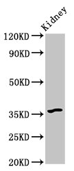
WB
Western Blot Positive WB detected in: Mouse kidney tissue All lanes: GPBAR1 antibody at 6µg/ml Secondary Goat polyclonal to rabbit IgG at 1/50000 dilution.IHC
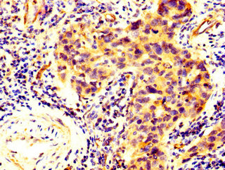
IHC image of GPBAR1 Antibody diluted at 1:300 and staining in paraffin-embedded human bladder cancer performed on a Leica BondTM system. After dewaxing and hydration, antigen retrieval was mediated by high pressure in a citrate buffer (pH 6.0). Section was blocked with 10% normal goat serum 30min at RT. Then primary antibody (1% BSA) was incubated at 4°C overnight. The primary is detected by a biotinylated secondary antibody and visualized using an HRP conjugated SP system.ICC/IF
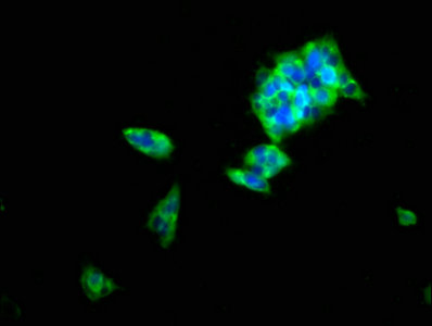
Immunofluorescence staining of 293 cells with GPBAR1 Antibody at 1:100, counter stained with DAPI. The cells were fixed in 4% formaldehyde, permeabilized using 0.2% Triton X-100 and blocked in 10% normal Goat Serum. The cells were then incubated with the antibody overnight at 4°C. The secondary antibody was Alexa Fluor 488-congugated AffiniPure Goat Anti-Rabbit IgG(H+L).| Product Name | Rabbit anti-GPBAR1 Polyclonal antibody |
|---|---|
| Antibody Type | Primary Antibodies |
| Immunogen | Recombinant Human G-protein coupled bile acid receptor 1 protein (283-330AA). |
| Clonality | Polyclonal |
|---|---|
| Isotype | IgG |
| Host Species | Rabbit |
| Tested Applications | ICC/IFIHCWB |
| WB:1:500-1:5000 IHC:1:100-1:300 ICC/IF:1:50-1:200 |
|
| Species Reactivity | HumanMouse |
| Concentration | 1mg/ml |
| Purification | Protein G |
| Gene Symbol | GPBAR1 |
|---|---|
| Gene Synonyms | BG37 TGR5 M-BAR GPCR19 GPR131 |
| Gene Full Name | G protein-coupled bile acid receptor 1 |
| Gene Summary | This gene encodes a member of the G protein-coupled receptor (GPCR) superfamily. This enzyme functions as a cell surface receptor for bile acids. Treatment of cells expressing this GPCR with bile acids induces the production of intracellular cAMP, activation of a MAP kinase signaling pathway, and internalization of the receptor. The receptor is implicated in the suppression of macrophage functions and regulation of energy homeostasis by bile acids. Alternative splicing results in multiple transcript variants encoding the same protein. [provided by RefSeq, Jul 2008] |
| Molecular Weight(MW) | 36kDa |
| Cellular Localization | Cell membrane. |
WB
Western Blot Positive WB detected in: Mouse kidney tissue All lanes: GPBAR1 antibody at 6µg/ml Secondary Goat polyclonal to rabbit IgG at 1/50000 dilution.
IHC
IHC image of GPBAR1 Antibody diluted at 1:300 and staining in paraffin-embedded human bladder cancer performed on a Leica BondTM system. After dewaxing and hydration, antigen retrieval was mediated by high pressure in a citrate buffer (pH 6.0). Section was blocked with 10% normal goat serum 30min at RT. Then primary antibody (1% BSA) was incubated at 4°C overnight. The primary is detected by a biotinylated secondary antibody and visualized using an HRP conjugated SP system.
ICC/IF
Immunofluorescence staining of 293 cells with GPBAR1 Antibody at 1:100, counter stained with DAPI. The cells were fixed in 4% formaldehyde, permeabilized using 0.2% Triton X-100 and blocked in 10% normal Goat Serum. The cells were then incubated with the antibody overnight at 4°C. The secondary antibody was Alexa Fluor 488-congugated AffiniPure Goat Anti-Rabbit IgG(H+L).| Application Notes | WB:1:500-1:5000 IHC:1:100-1:300 ICC/IF:1:50-1:200 |
|---|
| Form | Liquid |
|---|---|
| Storage Instructions | Upon receipt, store at -20°C or -80°C. Avoid repeated freeze. |
| Storage Buffer | Preservative: 0.03% Proclin 300,Constituents: 50% Glycerol, 0.01M PBS, pH 7.4 |


 Data sheet for OM644392