IHC
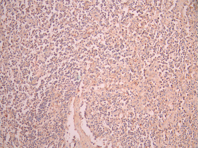
IHC image of CCL3 Antibody diluted at 1:50 and staining in paraffin-embedded human lymph node tissue performed on a Leica BondTM system. After dewaxing and hydration, antigen retrieval was mediated by high pressure in a citrate buffer (pH 6.0). Section was blocked with 10% normal goat serum 30min at RT. Then primary antibody (1% BSA) was incubated at 4°C overnight. The primary is detected by a Anti-Human lgG, Fcy Fragment Specific labeled by HRP and visualized using 0.05% DAB.IHC
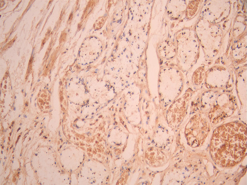
IHC image of CCL3 Antibody diluted at 1:50 and staining in paraffin-embedded human lung tissue performed on a Leica BondTM system. After dewaxing and hydration, antigen retrieval was mediated by high pressure in a citrate buffer (pH 6.0). Section was blocked with 10% normal goat serum 30min at RT. Then primary antibody (1% BSA) was incubated at 4°C overnight. The primary is detected by a Anti-Human lgG, Fcy Fragment Specific labeled by HRP and visualized using 0.05% DAB.FC
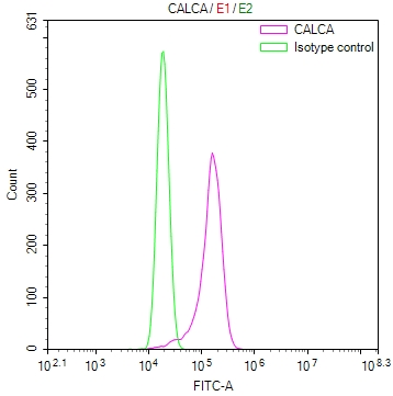
Overlay Peak curve showing K562 cells stained withCCL3 Antibody (red line) at 1:100. The cells were fixed in 4% formaldehyde and permeated by 0.2% TritonX-100 for 10min. Then 10% normal goat serum to block non-specific protein-protein interactions followed by the antibody (1ug/1*106cells) for 45min at 4℃. The secondary antibody used was Fluorescein (FITC) AffiniPure Goat Anti-Human IgG, Fcγ fragment specific at 1:200 dilution for 35min at 4℃.Control antibody (green line) was human IgG1 (1ug/1*106cells) used under the same conditions. Acquisition of >10,000 events was performed.| Product Name | CCL3 Recombinant Monoclonal Antibody |
|---|---|
| Antibody Type | Primary Antibodies |
| Immunogen | Recombinant Human CCL3 protein |
| Clonality | Monoclonal |
|---|---|
| Isotype | IgG1 |
| Tested Applications | FCIHC |
| IHC:1:50-1:200 FC:1:50-1:200 |
|
| Species Reactivity | Human |
| Concentration | 1mg/ml |
| Purification | Affinity purified |
| Gene Symbol | CCL3 |
|---|---|
| Gene Synonyms | SCI LD78 MIP1A SCYA3 G0S19-1 LD78ALPHA MIP-1-alpha |
| Gene Full Name | C-C motif chemokine ligand 3 |
| Gene Summary | This locus represents a small inducible cytokine. The encoded protein, also known as macrophage inflammatory protein 1 alpha, plays a role in inflammatory responses through binding to the receptors CCR1, CCR4 and CCR5. Polymorphisms at this locus may be associated with both resistance and susceptibility to infection by human immunodeficiency virus type 1.[provided by RefSeq, Sep 2010] |
| Alternative Names | C C motif chemokine 3 antibody CCL 3 antibody CCL3 antibody CCL3_HUMAN antibody Chemokine (C C motif) ligand 3 antibody Chemokine C C motif ligand 3 antibody Chemokine ligand 3 antibody G0/G1 switch regulatory protein 19 1 antibody G0/G1 switch regulatory protein 19-1 antibody G0S19 1 antibody G0S19 1 protein antibody Heparin binding chemotaxis protein antibody L2G25B antibody LD78 alpha antibody LD78-alpha(4-69) antibody LD78alpha antibody Macrophage inflammatory protein 1 alpha antibody Macrophage inflammatory protein 1-alpha antibody macrophage inflammatory protein 1a antibody MIP 1 alpha antibody MIP 1A antibody MIP-1-alpha antibody MIP-1-alpha(4-69) antibody MIP1 alpha antibody MIP1A antibody PAT 464.1 antibody SCYA 3 antibody SCYA3 antibody SIS alpha antibody SIS beta antibody SIS-beta antibody Small inducible cytokine A3 antibody small inducible cytokine A3 (homologous to mouse Mip-1a) antibody Small-inducible cytokine A3 antibody Tonsillar lymphocyte LD78 alpha protein antibody |
| Cellular Localization | Secreted. |
IHC
IHC image of CCL3 Antibody diluted at 1:50 and staining in paraffin-embedded human lymph node tissue performed on a Leica BondTM system. After dewaxing and hydration, antigen retrieval was mediated by high pressure in a citrate buffer (pH 6.0). Section was blocked with 10% normal goat serum 30min at RT. Then primary antibody (1% BSA) was incubated at 4°C overnight. The primary is detected by a Anti-Human lgG, Fcy Fragment Specific labeled by HRP and visualized using 0.05% DAB.
IHC
IHC image of CCL3 Antibody diluted at 1:50 and staining in paraffin-embedded human lung tissue performed on a Leica BondTM system. After dewaxing and hydration, antigen retrieval was mediated by high pressure in a citrate buffer (pH 6.0). Section was blocked with 10% normal goat serum 30min at RT. Then primary antibody (1% BSA) was incubated at 4°C overnight. The primary is detected by a Anti-Human lgG, Fcy Fragment Specific labeled by HRP and visualized using 0.05% DAB.
FC
Overlay Peak curve showing K562 cells stained withCCL3 Antibody (red line) at 1:100. The cells were fixed in 4% formaldehyde and permeated by 0.2% TritonX-100 for 10min. Then 10% normal goat serum to block non-specific protein-protein interactions followed by the antibody (1ug/1*106cells) for 45min at 4℃. The secondary antibody used was Fluorescein (FITC) AffiniPure Goat Anti-Human IgG, Fcγ fragment specific at 1:200 dilution for 35min at 4℃.Control antibody (green line) was human IgG1 (1ug/1*106cells) used under the same conditions. Acquisition of >10,000 events was performed.| Application Notes | IHC:1:50-1:200 FC:1:50-1:200 |
|---|
| Form | Liquid |
|---|---|
| Storage Instructions | Upon receipt, store at -20°C or -80°C. Avoid repeated freeze. |
| Storage Buffer | 0.03% Proclin 300 Constituents: 50% Glycerol, 0.01M PBS, PH 7.4 |


