WB
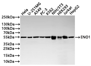
Western Blot: Positive WB detected in: HeLa whole cell lysate(20µg), U-251MG whole cell lysate(20µg), A549 whole cell lysate(20µg), PC-3 whole cell lysate(20µg), K562 whole cell lysate(20µg), NIH/3T3 whole cell lysate(20µg), HEK293 whole cell lysate(20µg), Jurkat whole cell lysate(20µg), HepG2 whole cell lysate(20µg). All lanes: ENO1 antibody at 1:1000. Secondary: Goat polyclonal to human IgG at 1/40000 dilution. Exposure time:5sWB
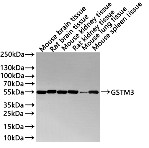
Western Blot: Positive WB detected in: Mouse brain tissue lysate(20µg), Rat brain tissue lysate(20µg), Mouse kidney tissue lysate(20µg), Rat kidney tissue lysate(20µg), Mouse lung tissue lysate(20µg), Mouse spleen tissue lysate(20µg). All lanes: ENO1 antibody at 1:1000. Secondary: Goat polyclonal to human IgG at 1/40000 dilution.IHC
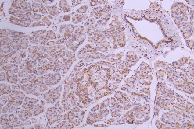
IHC image of ENO1 Recombinant Monoclonal Antibody diluted at 1:50 and staining in paraffin-embedded human pancreatic tissue performed on a Leica BondTM system. After dewaxing and hydration, antigen retrieval was mediated by high pressure in a citrate buffer (pH 6.0). Section was blocked with 10% normal goat serum 30min at RT. Then primary antibody (1% BSA) was incubated at 4°C overnight. The primary is detected by a Goat anti-human polymer IgG labeled by HRP and visualized using 0.05% DAB.ICC/IF
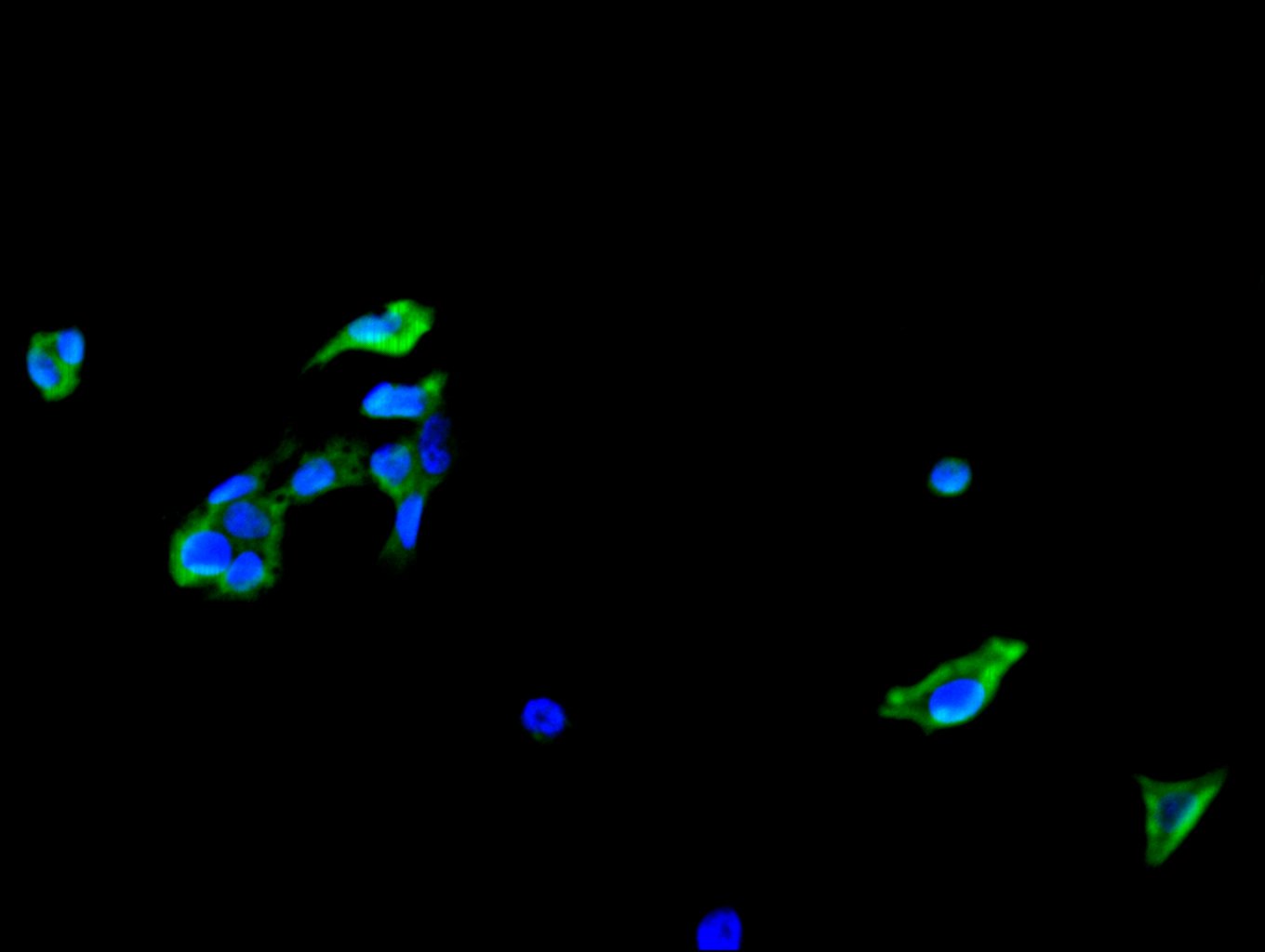
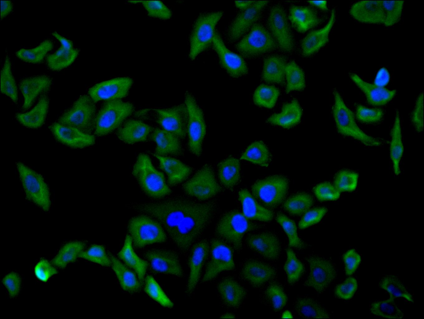
Immunofluorescence staining of HepG2 cell with ENO1 Recombinant Monoclonal Antibody at 1:30 counter-stained with DAPI. The cells were fixed in 4% formaldehyde, permeabilized using 0.2% Triton X-100 and blocked in 10% normal Goat Serum. The cells were then incubated with the antibody overnight at 4°C. The secondary antibody was Fluorescein (FITC) AffiniPure Goat Anti-Human IgG, Fcγ fragment specific.ICC/IF
Immunofluorescence staining of MCF-7 cell with ENO1 Recombinant Monoclonal Antibody at 1:30 counter-stained with DAPI. The cells were fixed in 4% formaldehyde, permeabilized using 0.2% Triton X-100 and blocked in 10% normal Goat Serum. The cells were then incubated with the antibody overnight at 4°C. The secondary antibody was Fluorescein (FITC) AffiniPure Goat Anti-Human IgG, Fcγ fragment specific.FC
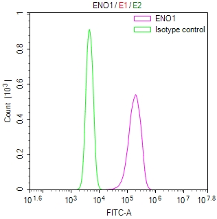
Overlay Peak curve showing Hela cells stained with ENO1 Recombinant Monoclonal Antibody (red line) at 1:100. The cells were fixed in 4% formaldehyde and permeated by 0.2% TritonX-100 for 10min. Then 10% normal goat serum to block non-specific protein-protein interactions followed by the antibody (1ug/1*106cells) for 45min at 4℃. The secondary antibody used was Fluorescein (FITC) AffiniPure Goat Anti-Human IgG, Fcγ fragment specific at 1:200 dilution for 35min at 4℃.Control antibody (green line) was human IgG1 (1ug/1*106cells) used under the same conditions. Acquisition of >10,000 events was performed.| Product Name | ENO1 Recombinant Monoclonal Antibody |
|---|---|
| Antibody Type | Primary Antibodies |
| Immunogen | Recombinant Human ENO1 protein |
| Clonality | Monoclonal |
|---|---|
| Isotype | IgG1 |
| Host Species | Human |
| Tested Applications | FCICC/IFIHCWB |
| WB:1:500-1:2000 IHC:1:50-1:200 ICC/IF:1:50-1:200 FC:1:50-1:200 |
|
| Species Reactivity | HumanMouseRat |
| Concentration | 1mg/ml |
| Purification | Affinity purified |
| Gene Symbol | ENO1 |
|---|---|
| Gene Synonyms | NNE PPH MPB1 ENO1L1 ENO1-IT1 HEL-S-17 |
| Gene Full Name | enolase 1 |
| Gene Summary | This gene encodes alpha-enolase, one of three enolase isoenzymes found in mammals. Each isoenzyme is a homodimer composed of 2 alpha, 2 gamma, or 2 beta subunits, and functions as a glycolytic enzyme. Alpha-enolase in addition, functions as a structural lens protein (tau-crystallin) in the monomeric form. Alternative splicing of this gene results in a shorter isoform that has been shown to bind to the c-myc promoter and function as a tumor suppressor. Several pseudogenes have been identified, including one on the long arm of chromosome 1. Alpha-enolase has also been identified as an autoantigen in Hashimoto encephalopathy. [provided by RefSeq, Jan 2011] |
| Alternative Names | 2 phospho D glycerate hydro lyase antibody 2-phospho-D-glycerate hydro-lyase antibody Alpha enolase antibody Alpha enolase like 1 antibody Alpha-enolase antibody C myc promoter binding protein antibody C-myc promoter-binding protein antibody EC 4.2.1.11 antibody eno1 antibody ENO1L1 antibody ENOA_HUMAN antibody Enolase 1 (alpha) antibody Enolase 1 (alpha) like 1 antibody Enolase 1 antibody Enolase alpha antibody MBP 1 antibody MBP-1 antibody MBP1 antibody MBPB1 antibody MPB 1 antibody MPB-1 antibody MPB1 antibody MYC promoter binding protein 1 antibody NNE antibody Non neural enolase antibody Non-neural enolase antibody Phosphopyruvate hydratase antibody Plasminogen binding protein antibody Plasminogen-binding protein antibody PPH antibody Tau crystallin antibody |
| Molecular Weight(MW) | 47 kDa(Observed band size: 55 kDa) |
| Cellular Localization | Cytoplasm. Cell membrane. Cytoplasm, myofibril, sarcomere, M line. Note=Can translocate to the plasma membrane in either the homodimeric (alpha/alpha) or heterodimeric (alpha/gamma) form. ENO1 is localized to the M line.; [Isoform MBP-1]: Nucleus. |
WB
Western Blot: Positive WB detected in: HeLa whole cell lysate(20µg), U-251MG whole cell lysate(20µg), A549 whole cell lysate(20µg), PC-3 whole cell lysate(20µg), K562 whole cell lysate(20µg), NIH/3T3 whole cell lysate(20µg), HEK293 whole cell lysate(20µg), Jurkat whole cell lysate(20µg), HepG2 whole cell lysate(20µg). All lanes: ENO1 antibody at 1:1000. Secondary: Goat polyclonal to human IgG at 1/40000 dilution. Exposure time:5s
WB
Western Blot: Positive WB detected in: Mouse brain tissue lysate(20µg), Rat brain tissue lysate(20µg), Mouse kidney tissue lysate(20µg), Rat kidney tissue lysate(20µg), Mouse lung tissue lysate(20µg), Mouse spleen tissue lysate(20µg). All lanes: ENO1 antibody at 1:1000. Secondary: Goat polyclonal to human IgG at 1/40000 dilution.
IHC
IHC image of ENO1 Recombinant Monoclonal Antibody diluted at 1:50 and staining in paraffin-embedded human pancreatic tissue performed on a Leica BondTM system. After dewaxing and hydration, antigen retrieval was mediated by high pressure in a citrate buffer (pH 6.0). Section was blocked with 10% normal goat serum 30min at RT. Then primary antibody (1% BSA) was incubated at 4°C overnight. The primary is detected by a Goat anti-human polymer IgG labeled by HRP and visualized using 0.05% DAB.
ICC/IF
Immunofluorescence staining of HepG2 cell with ENO1 Recombinant Monoclonal Antibody at 1:30 counter-stained with DAPI. The cells were fixed in 4% formaldehyde, permeabilized using 0.2% Triton X-100 and blocked in 10% normal Goat Serum. The cells were then incubated with the antibody overnight at 4°C. The secondary antibody was Fluorescein (FITC) AffiniPure Goat Anti-Human IgG, Fcγ fragment specific.
ICC/IF
Immunofluorescence staining of MCF-7 cell with ENO1 Recombinant Monoclonal Antibody at 1:30 counter-stained with DAPI. The cells were fixed in 4% formaldehyde, permeabilized using 0.2% Triton X-100 and blocked in 10% normal Goat Serum. The cells were then incubated with the antibody overnight at 4°C. The secondary antibody was Fluorescein (FITC) AffiniPure Goat Anti-Human IgG, Fcγ fragment specific.
FC
Overlay Peak curve showing Hela cells stained with ENO1 Recombinant Monoclonal Antibody (red line) at 1:100. The cells were fixed in 4% formaldehyde and permeated by 0.2% TritonX-100 for 10min. Then 10% normal goat serum to block non-specific protein-protein interactions followed by the antibody (1ug/1*106cells) for 45min at 4℃. The secondary antibody used was Fluorescein (FITC) AffiniPure Goat Anti-Human IgG, Fcγ fragment specific at 1:200 dilution for 35min at 4℃.Control antibody (green line) was human IgG1 (1ug/1*106cells) used under the same conditions. Acquisition of >10,000 events was performed.| Application Notes | WB:1:500-1:2000 IHC:1:50-1:200 ICC/IF:1:50-1:200 FC:1:50-1:200 |
|---|
| Form | Liquid |
|---|---|
| Storage Instructions | Upon receipt, store at -20°C or -80°C. Avoid repeated freeze. |
| Storage Buffer | 0.03% Proclin 300 Constituents: 50% Glycerol, 0.01M PBS, PH 7.4 |


