IF-F
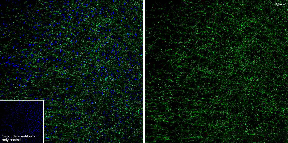
Application: Immunofluorescence (IHC-Fr). Species: Mouse. Tissue: Cerebral cortex. Sample: Frozen section. Antigen retrieval: Not required. Wash buffer: 1× PBS. Blocking: 10% normal goat serum + 0.5 % Triton X-100 + 0.3 M Glycine in PBS, 10 minutes at room temperature. Primary antibody: 1/500, overnight at 4°C. Secondary antibody: Goat Anti-Rat IgG (488), 1.5 hours at room temperature.IHC
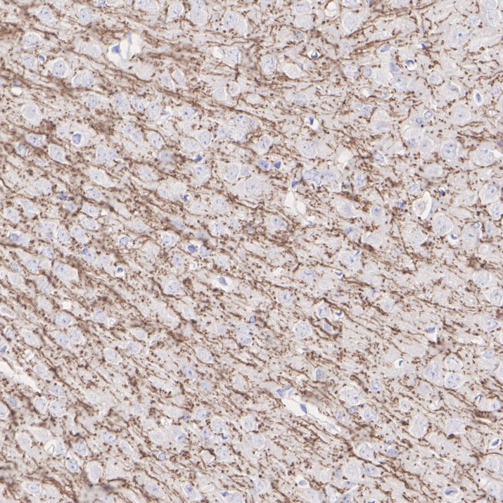
Application: Immunohistochemistry (IHC-P) . Species: Mouse . Tissue: Cerebral cortex. Sample: Paraffin-embedded section . Antigen retrieval: Heat-mediated, Tris-EDTA buffer (pH 9.0), 20 minutes at 95°C. Wash buffer: 1× TBST . Endogenous peroxidase blocking: 3% H2O2, 10 minutes at room temperature. Blocking: 1% BSA + 10% normal goat serum, 10 minutes at room temperature. Primary antibody: 1/5,000, 1 hour at room temperature. Secondary antibody:30 minutes at room temperature.IHC
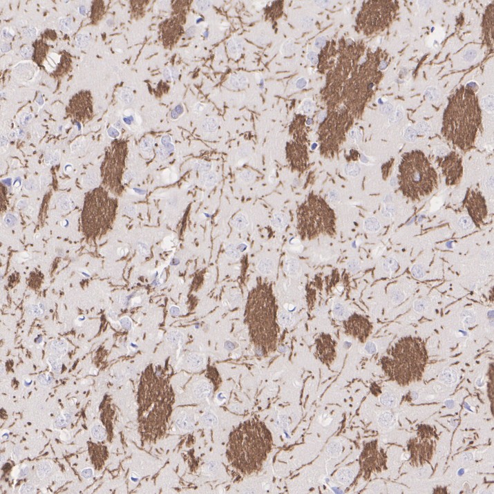
Application: Immunohistochemistry (IHC-P) . Species: Rat. Tissue: Brain. Sample: Paraffin-embedded section . Antigen retrieval: Heat-mediated, Tris-EDTA buffer (pH 9.0), 20 minutes at 95°C. Wash buffer: 1× TBST . Endogenous peroxidase blocking: 3% H2O2, 10 minutes at room temperature. Blocking: 1% BSA + 10% normal goat serum, 10 minutes at room temperature. Primary antibody:1/5,000, 1 hour at room temperature. Secondary antibody:30 minutes at room temperature.WB
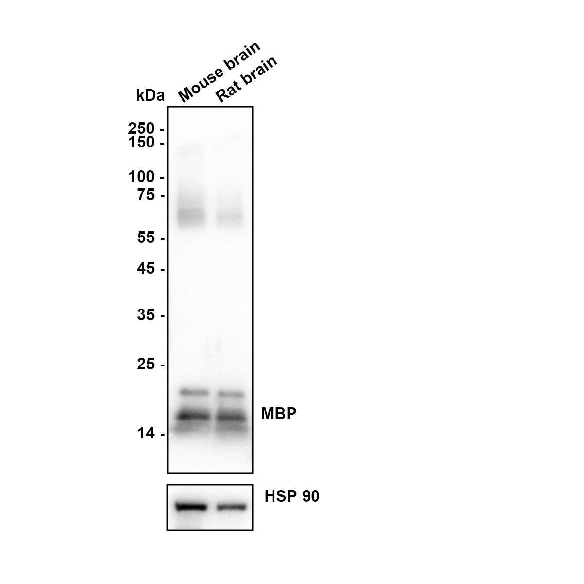
Western blot analysis of Myelin Basic Protein on different lysates with Rat anti-Myelin Basic Protein antibody at 1/5,000 dilution. Lysates/proteins at 20 µg/Lane. Exposure time: 8 seconds. Blocking: 5% NFDM/TBST, 1 hour at room temperature. Primary antibody:1/5,000 in primary antibody dilution buffer, overnight at 4 ℃. Secondary antibody: Goat anti-Rat IgG-HRP, 1/50,000 in 5% NFDM/TBST, 1 hour at room temperature.| Product Name | Myelin Basic Protein Recombinant Antibody - Rat IgG1 (Chimeric) |
|---|---|
| Antibody Type | Primary Antibodies |
| Immunogen | Recombinant protein within Human Myelin Basic Protein aa 121-304 / 304. |
| Clonality | Monoclonal |
|---|---|
| Isotype | IgG1 |
| Tested Applications | IF-FIHCWB |
| IF-F:1:500 IHC:1:5000 WB:1:5000 |
|
| Species Reactivity | MouseRat |
| Concentration | 1mg/ml |
| Purification | Protein A |
| Gene Symbol | Mbp |
|---|---|
| Gene Synonyms | jve mld shi Hmbpr golli-mbp |
| Gene Full Name | myelin basic protein |
| Gene Summary | The protein encoded by the classic Mbp gene is a major constituent of the myelin sheath of oligodendrocytes and Schwann cells in the nervous system. However, Mbp-related transcripts are also present in the bone marrow and the immune system. These mRNAs arise from the long Mbp gene (otherwise called "Golli-Mbp") that contains 3 additional exons located upstream of the classic Mbp exons. Alternative splicing from the Golli and the Mbp transcription start sites gives rise to 2 sets of Mbp-related transcripts and gene products. The Golli mRNAs contain 3 exons unique to Golli-Mbp, spliced in-frame to 1 or more Mbp exons. They encode hybrid proteins that have N-terminal Golli aa sequence linked to Mbp aa sequence. The second family of transcripts contain only Mbp exons and produce the well characterized myelin basic proteins. This complex gene structure is conserved among species suggesting that the Mbp transcription unit is an integral part of the Golli transcription unit and that this arrangement is important for the function and/or regulation of these genes. Mutation of the Mbp gene is associated with the 'shiverer' and 'myelin deficient' phenotypes in mouse. [provided by RefSeq, Jul 2008] |
| Alternative Names | GDB antibody Golli MBP antibody Golli MBP myelin basic protein antibody Hemopoietic MBP antibody HMBPR antibody HUGO antibody MBP antibody MBP_CAVPO antibody MBP_HUMAN antibody GDB antibody Golli MBP antibody Golli MBP myelin basic protein antibody Hemopoietic MBP antibody HMBPR antibody HUGO antibody MBP antibody MBP_CAVPO antibody MBP_HUMAN antibody MGC99675 antibody MLD antibody Myelin A1 protein antibody Myelin A1 Protein basic antibody Myelin basic protein antibody Myelin Deficient antibody Myelin membrane encephalitogenic protein antibody OTTHUMP00000163776 antibody OTTHUMP00000174387 antibody OTTHUMP00000174388 antibody SHI antibody Shiverer antibody SP antibody |
| Molecular Weight(MW) | 33 kDa(Observed band size: 14-20 kDa) |
| Cellular Localization | Myelin membrane, Nucleus. |
IF-F
Application: Immunofluorescence (IHC-Fr). Species: Mouse. Tissue: Cerebral cortex. Sample: Frozen section. Antigen retrieval: Not required. Wash buffer: 1× PBS. Blocking: 10% normal goat serum + 0.5 % Triton X-100 + 0.3 M Glycine in PBS, 10 minutes at room temperature. Primary antibody: 1/500, overnight at 4°C. Secondary antibody: Goat Anti-Rat IgG (488), 1.5 hours at room temperature.
IHC
Application: Immunohistochemistry (IHC-P) . Species: Mouse . Tissue: Cerebral cortex. Sample: Paraffin-embedded section . Antigen retrieval: Heat-mediated, Tris-EDTA buffer (pH 9.0), 20 minutes at 95°C. Wash buffer: 1× TBST . Endogenous peroxidase blocking: 3% H2O2, 10 minutes at room temperature. Blocking: 1% BSA + 10% normal goat serum, 10 minutes at room temperature. Primary antibody: 1/5,000, 1 hour at room temperature. Secondary antibody:30 minutes at room temperature.
IHC
Application: Immunohistochemistry (IHC-P) . Species: Rat. Tissue: Brain. Sample: Paraffin-embedded section . Antigen retrieval: Heat-mediated, Tris-EDTA buffer (pH 9.0), 20 minutes at 95°C. Wash buffer: 1× TBST . Endogenous peroxidase blocking: 3% H2O2, 10 minutes at room temperature. Blocking: 1% BSA + 10% normal goat serum, 10 minutes at room temperature. Primary antibody:1/5,000, 1 hour at room temperature. Secondary antibody:30 minutes at room temperature.
WB
Western blot analysis of Myelin Basic Protein on different lysates with Rat anti-Myelin Basic Protein antibody at 1/5,000 dilution. Lysates/proteins at 20 µg/Lane. Exposure time: 8 seconds. Blocking: 5% NFDM/TBST, 1 hour at room temperature. Primary antibody:1/5,000 in primary antibody dilution buffer, overnight at 4 ℃. Secondary antibody: Goat anti-Rat IgG-HRP, 1/50,000 in 5% NFDM/TBST, 1 hour at room temperature.| Application Notes | IF-F:1:500 IHC:1:5000 WB:1:5000 |
|---|
| Form | Liquid |
|---|---|
| Storage Instructions | Shipped at 4℃. Store at +4℃ short term (1-2 weeks). It is recommended to aliquot into single-use upon delivery. Store at -20℃ long term. |
| Storage Buffer | 1*PBS (pH7.4), 0.1% BSA, 40% Glycerol, 0.2% Proclean 950. |


