IF-F
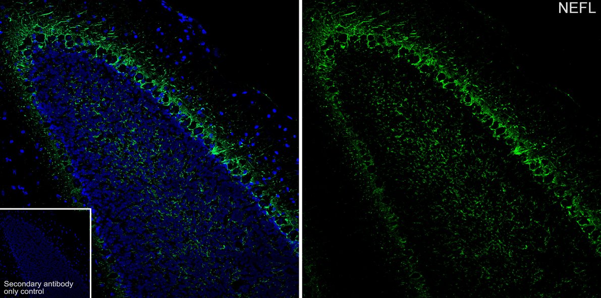
Application: IHC-Fr. Species: Mouse. Site: Cerebellum. Sample: Frozen section. Antibody concentration: 1:500. Antigen retrieval: Not required.IHC
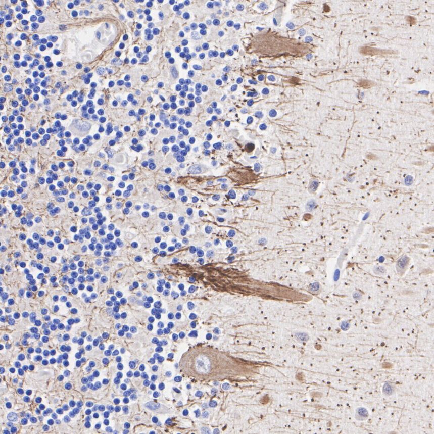
Immunohistochemical analysis of paraffin-embedded human cerebellum tissue with Rat anti-NF-L antibody at 1/2,000 dilution. The section was pre-treated using heat mediated antigen retrieval with Tris-EDTA buffer (pH 9.0) for 20 minutes. The tissues were blocked in 1% BSA for 20 minutes at room temperature, washed with ddH2O and PBS, and then probed with the primary antibody at 1/2,000 dilution for 1 hour at room temperature. The detection was performed using an HRP conjugated compact polymer system. DAB was used as the chromogen. Tissues were counterstained with hematoxylin and mounted with DPX.IHC
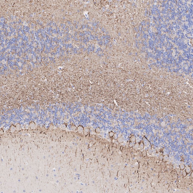
Immunohistochemical analysis of paraffin-embedded mouse cerebellum tissue with Rat anti-NF-L antibody at 1/4,000 dilution. The section was pre-treated using heat mediated antigen retrieval with Tris-EDTA buffer (pH 9.0) for 20 minutes. The tissues were blocked in 1% BSA for 20 minutes at room temperature, washed with ddH2O and PBS, and then probed with the primary antibody at 1/4,000 dilution for 1 hour at room temperature. The detection was performed using an HRP conjugated compact polymer system. DAB was used as the chromogen. Tissues were counterstained with hematoxylin and mounted with DPX.IHC
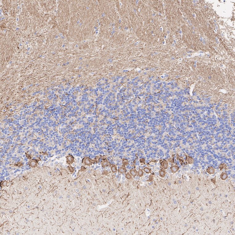
Immunohistochemical analysis of paraffin-embedded rat cerebellum tissue with Rat anti-NF-L antibody at 1/4,000 dilution. The section was pre-treated using heat mediated antigen retrieval with Tris-EDTA buffer (pH 9.0) for 20 minutes. The tissues were blocked in 1% BSA for 20 minutes at room temperature, washed with ddH2O and PBS, and then probed with the primary antibody at 1/4,000 dilution for 1 hour at room temperature. The detection was performed using an HRP conjugated compact polymer system. DAB was used as the chromogen. Tissues were counterstained with hematoxylin and mounted with DPX.WB
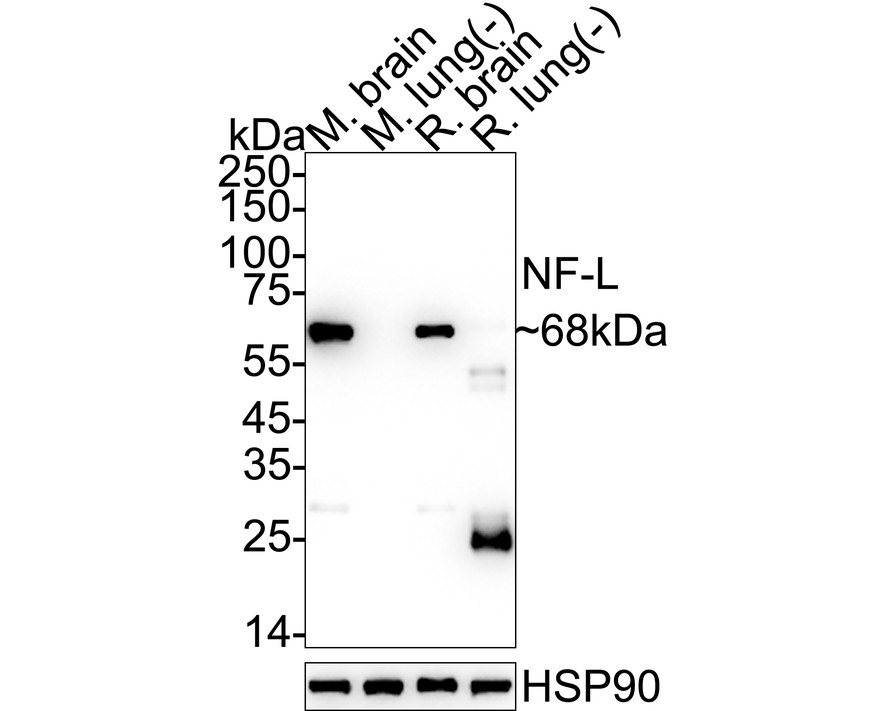
Western blot analysis of NF-L on different lysates with Rat anti-NF-L antibody at 1/2,000 dilution. Lysates/proteins at 20 µg/Lane. Exposure time: 10 seconds; 4-20% SDS-PAGE gel. Proteins were transferred to a PVDF membrane and blocked with 5% NFDM/TBST for 1 hour at room temperature. The primary antibody at 1/2,000 dilution was used in 5% NFDM/TBST at 4℃ overnight. Goat Anti-Rat IgG H&L - HRP Secondary Antibody at 1/5,000 dilution was used for 1 hour at room temperature.| Product Name | NF-L Recombinant Antibody - Rat IgG1 (Chimeric) |
|---|---|
| Antibody Type | Primary Antibodies |
| Immunogen | Recombinant protein. |
| Isotype | IgG1 |
|---|---|
| Host Species | Rat |
| Tested Applications | IF-FIHCWB |
| WB:1:2000 IHC:1:2000-1:4000 IF-F:1:500 |
|
| Species Reactivity | HumanMouseRat |
| Concentration | 1mg/ml |
| Purification | Protein A |
| Gene Symbol | NEFL |
|---|---|
| Gene Synonyms | NFL NF-L NF68 CMT1F CMT2E CMTDIG PPP1R110 |
| Gene Full Name | neurofilament light chain |
| Gene Summary | Neurofilaments are type IV intermediate filament heteropolymers composed of light, medium, and heavy chains. Neurofilaments comprise the axoskeleton and they functionally maintain the neuronal caliber. They may also play a role in intracellular transport to axons and dendrites. This gene encodes the light chain neurofilament protein. Mutations in this gene cause Charcot-Marie-Tooth disease types 1F (CMT1F) and 2E (CMT2E), disorders of the peripheral nervous system that are characterized by distinct neuropathies. A pseudogene has been identified on chromosome Y. [provided by RefSeq, Oct 2008] |
| Alternative Names | 68 kDa neurofilament protein antibody 68kDa Neurofilament antibody 68kDa neurofilament protein antibody CMT1F antibody CMT2E antibody FLJ53642 antibody Light molecular weight neurofilament protein antibody NEFL antibody Neurofilament light antibody Neurofilament light polypeptide 68kDa antibody 68 kDa neurofilament protein antibody 68kDa Neurofilament antibody 68kDa neurofilament protein antibody CMT1F antibody CMT2E antibody FLJ53642 antibody Light molecular weight neurofilament protein antibody NEFL antibody Neurofilament light antibody Neurofilament light polypeptide 68kDa antibody Neurofilament light polypeptide antibody Neurofilament protein light chain antibody Neurofilament subunit NF L antibody Neurofilament triplet L protein antibody NF-L antibody NF68 antibody NFL antibody NFL_HUMAN antibody |
| Molecular Weight(MW) | 62 kDa(Observed band size: 68 kDa) |
| Cellular Localization | Cell projection, axon, Cytoplasm, cytoskeleton. |
IF-F
Application: IHC-Fr. Species: Mouse. Site: Cerebellum. Sample: Frozen section. Antibody concentration: 1:500. Antigen retrieval: Not required.
IHC
Immunohistochemical analysis of paraffin-embedded human cerebellum tissue with Rat anti-NF-L antibody at 1/2,000 dilution. The section was pre-treated using heat mediated antigen retrieval with Tris-EDTA buffer (pH 9.0) for 20 minutes. The tissues were blocked in 1% BSA for 20 minutes at room temperature, washed with ddH2O and PBS, and then probed with the primary antibody at 1/2,000 dilution for 1 hour at room temperature. The detection was performed using an HRP conjugated compact polymer system. DAB was used as the chromogen. Tissues were counterstained with hematoxylin and mounted with DPX.
IHC
Immunohistochemical analysis of paraffin-embedded mouse cerebellum tissue with Rat anti-NF-L antibody at 1/4,000 dilution. The section was pre-treated using heat mediated antigen retrieval with Tris-EDTA buffer (pH 9.0) for 20 minutes. The tissues were blocked in 1% BSA for 20 minutes at room temperature, washed with ddH2O and PBS, and then probed with the primary antibody at 1/4,000 dilution for 1 hour at room temperature. The detection was performed using an HRP conjugated compact polymer system. DAB was used as the chromogen. Tissues were counterstained with hematoxylin and mounted with DPX.
IHC
Immunohistochemical analysis of paraffin-embedded rat cerebellum tissue with Rat anti-NF-L antibody at 1/4,000 dilution. The section was pre-treated using heat mediated antigen retrieval with Tris-EDTA buffer (pH 9.0) for 20 minutes. The tissues were blocked in 1% BSA for 20 minutes at room temperature, washed with ddH2O and PBS, and then probed with the primary antibody at 1/4,000 dilution for 1 hour at room temperature. The detection was performed using an HRP conjugated compact polymer system. DAB was used as the chromogen. Tissues were counterstained with hematoxylin and mounted with DPX.
WB
Western blot analysis of NF-L on different lysates with Rat anti-NF-L antibody at 1/2,000 dilution. Lysates/proteins at 20 µg/Lane. Exposure time: 10 seconds; 4-20% SDS-PAGE gel. Proteins were transferred to a PVDF membrane and blocked with 5% NFDM/TBST for 1 hour at room temperature. The primary antibody at 1/2,000 dilution was used in 5% NFDM/TBST at 4℃ overnight. Goat Anti-Rat IgG H&L - HRP Secondary Antibody at 1/5,000 dilution was used for 1 hour at room temperature.| Application Notes | WB:1:2000 IHC:1:2000-1:4000 IF-F:1:500 |
|---|
| Form | Liquid |
|---|---|
| Storage Instructions | Shipped at 4℃. Store at +4℃ short term (1-2 weeks). Store at -20℃ long term. |
| Storage Buffer | 1*PBS (pH7.4), 0.1% BSA, 40% Glycerol, 0.2% Proclean 950. |


