IHC
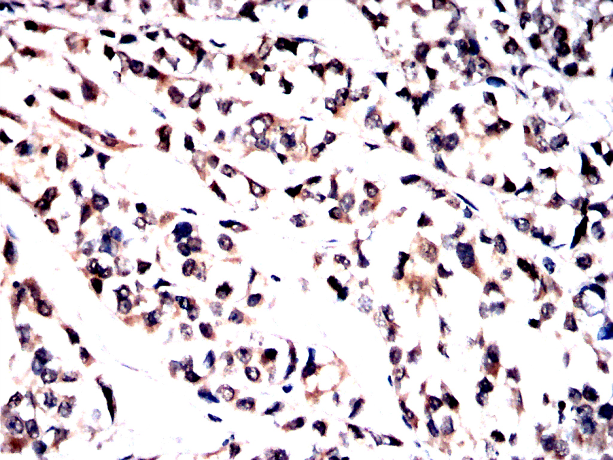
Immunohistochemical analysis of paraffin-embedded Human bladder cancer tissues using Tyk2 antibody with DAB staining.IHC
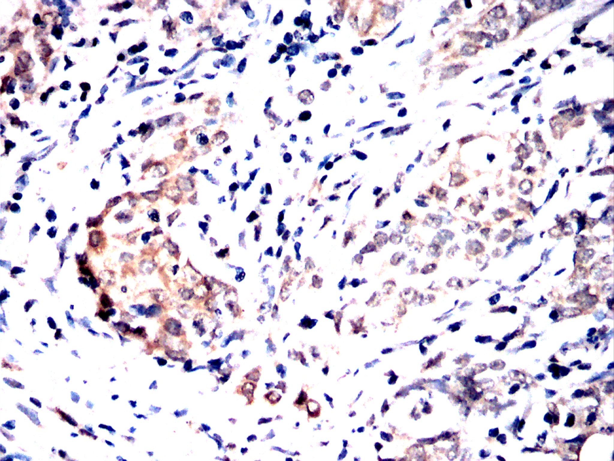
Immunohistochemical analysis of paraffin-embedded Human cervical cancer tissues using Tyk2 antibody with DAB staining.IHC
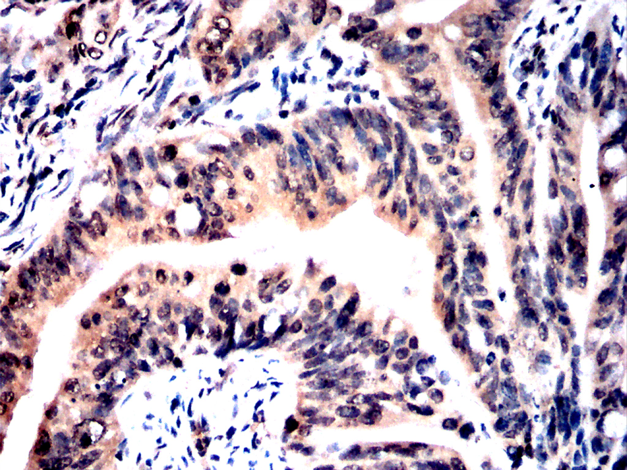
Immunohistochemical analysis of paraffin-embedded Human rectal cancer tissues using Tyk2 antibody with DAB staining.IHC
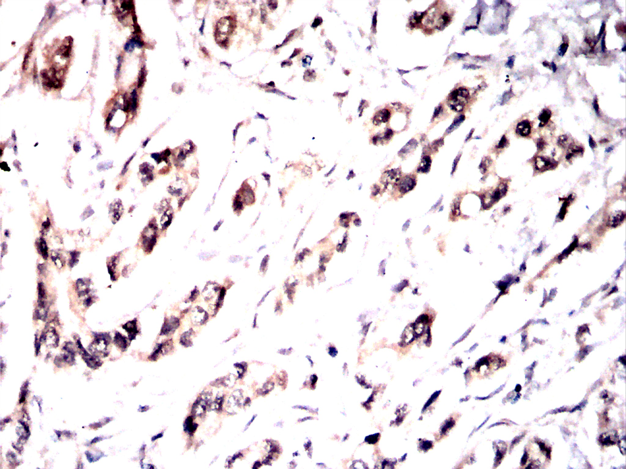
Immunohistochemical analysis of paraffin-embedded Human stomach cancer tissues using Tyk2 antibody with DAB staining.WB
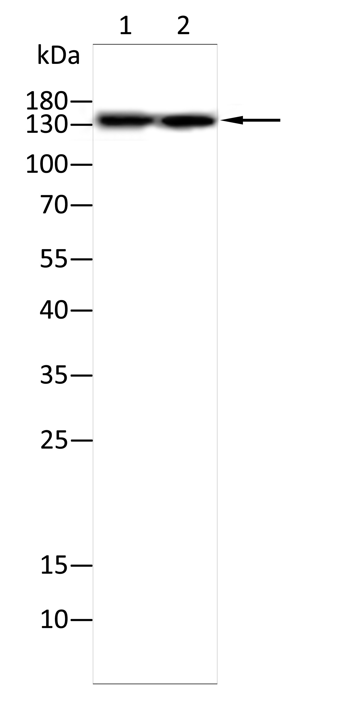
Western blot analysis using Tyk2 antibody against A431(1),Raji(2) cell .10% SDS-PAGE gel.Sample loading: 20μg /lane. Transfer the proteins onto a PVDF membrane (OM790003), and block it with TBST (OM750016) plus skimmed milk powder(OM750084) for one hour. Dilute the primary antibody with the antibody diluent (OM750012) at a ratio of 1:2000, and incubate it overnight at 4°C. Wash the membrane three times with TBST (OM750016), 5 minutes each time. At room temperature, dilute the secondary antibody, Goat Anti-Rabbit IgG(H&L)-HRP (OM643487), at a ratio of 1:20000 and incubate for one hour. Wash the membrane three times with TBST (OM750016) again, 5 minutes each time. Use ECL (OM625701) for luminescence.| Product Name | Anti-Tyk2 antibody-HRP |
|---|---|
| Antibody Type | Primary Antibodies |
| Immunogen | Polypeptide |
| Conjugation | HRP |
| Clonality | Polyclonal |
|---|---|
| Isotype | IgG |
| Host Species | Rabbit |
| Tested Applications | WBIHC |
| WB:1:200-1:2000 IHC:1:100-1:1000 |
|
| Species Reactivity | HumanMouseRat |
| Concentration | 1mg/ml |
| Purification | Protein A |
| Gene Symbol | TYK2 |
|---|---|
| Gene Synonyms | JTK1 IMD35 |
| Gene Full Name | tyrosine kinase 2 |
| Gene Summary | This gene encodes a member of the tyrosine kinase and, more specifically, the Janus kinases (JAKs) protein families. This protein associates with the cytoplasmic domain of type I and type II cytokine receptors and promulgate cytokine signals by phosphorylating receptor subunits. It is also a component of both the type I and type III interferon signaling pathways. As such, it may play a role in anti-viral immunity. A mutation in this gene has been associated with Immunodeficiency 35. [provided by RefSeq, Sep 2020] |
| Molecular Weight(MW) | 134kDa |
| Source | Rabbit |
| Cellular Localization | Cytoskeleton,Nucleus,Cytosol,Plasma membrane |
| SwissProt ID | P29597 |
|---|
IHC
Immunohistochemical analysis of paraffin-embedded Human bladder cancer tissues using Tyk2 antibody with DAB staining.
IHC
Immunohistochemical analysis of paraffin-embedded Human cervical cancer tissues using Tyk2 antibody with DAB staining.
IHC
Immunohistochemical analysis of paraffin-embedded Human rectal cancer tissues using Tyk2 antibody with DAB staining.
IHC
Immunohistochemical analysis of paraffin-embedded Human stomach cancer tissues using Tyk2 antibody with DAB staining.
WB
Western blot analysis using Tyk2 antibody against A431(1),Raji(2) cell .10% SDS-PAGE gel.Sample loading: 20μg /lane. Transfer the proteins onto a PVDF membrane (OM790003), and block it with TBST (OM750016) plus skimmed milk powder(OM750084) for one hour. Dilute the primary antibody with the antibody diluent (OM750012) at a ratio of 1:2000, and incubate it overnight at 4°C. Wash the membrane three times with TBST (OM750016), 5 minutes each time. At room temperature, dilute the secondary antibody, Goat Anti-Rabbit IgG(H&L)-HRP (OM643487), at a ratio of 1:20000 and incubate for one hour. Wash the membrane three times with TBST (OM750016) again, 5 minutes each time. Use ECL (OM625701) for luminescence.| Application Notes | WB:1:200-1:2000 IHC:1:100-1:1000 |
|---|
| Form | Liquid |
|---|---|
| Storage Instructions | Shipped at 4°C. Store at +4°C short term (1-2 weeks). Store at -20°C long term. Avoid freeze / thaw cycle. |
| Storage Buffer | Purified antibody in PBS with 0.05% sodium azide. |


