ICC/IF
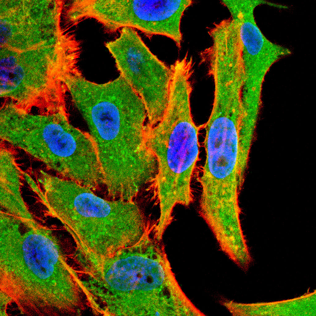
Immunofluorescence analysis of Hela cells using β-Actin antibody (green). Blue: DAPI fluorescent DNA dye. Red: Actin filaments have been labeled with Omnimabs® 594-Phalloidin.Cells are fixed in 4% paraformaldehyde at room temperature for 15 minutes. Then, they are permeabilized with a PBS (OM750003) solution containing 0.1% Triton X-100(OM750021) at room temperature for 15 minutes. Subsequently, the cells are blocked with 10% non - immune goat serum(OM760028) at room temperature for 1 hour.The cells are incubated overnight at 4°C with the primary antibody diluted 1:100 in PBS. The secondary antibody, Omnimabs® 488 Goat Ant-Rabbit IgG(H&L) (Green,OM643486), is diluted at a ratio of 1:400 and incubated with the cells for 1 hour.Nuclear DNA is labeled with DAPI (Blue,OM643160). F-actin is stained with Omnimabs® 594-Phalloidin (Red,OM750007) diluted 1:100 for 30 minutes.WB
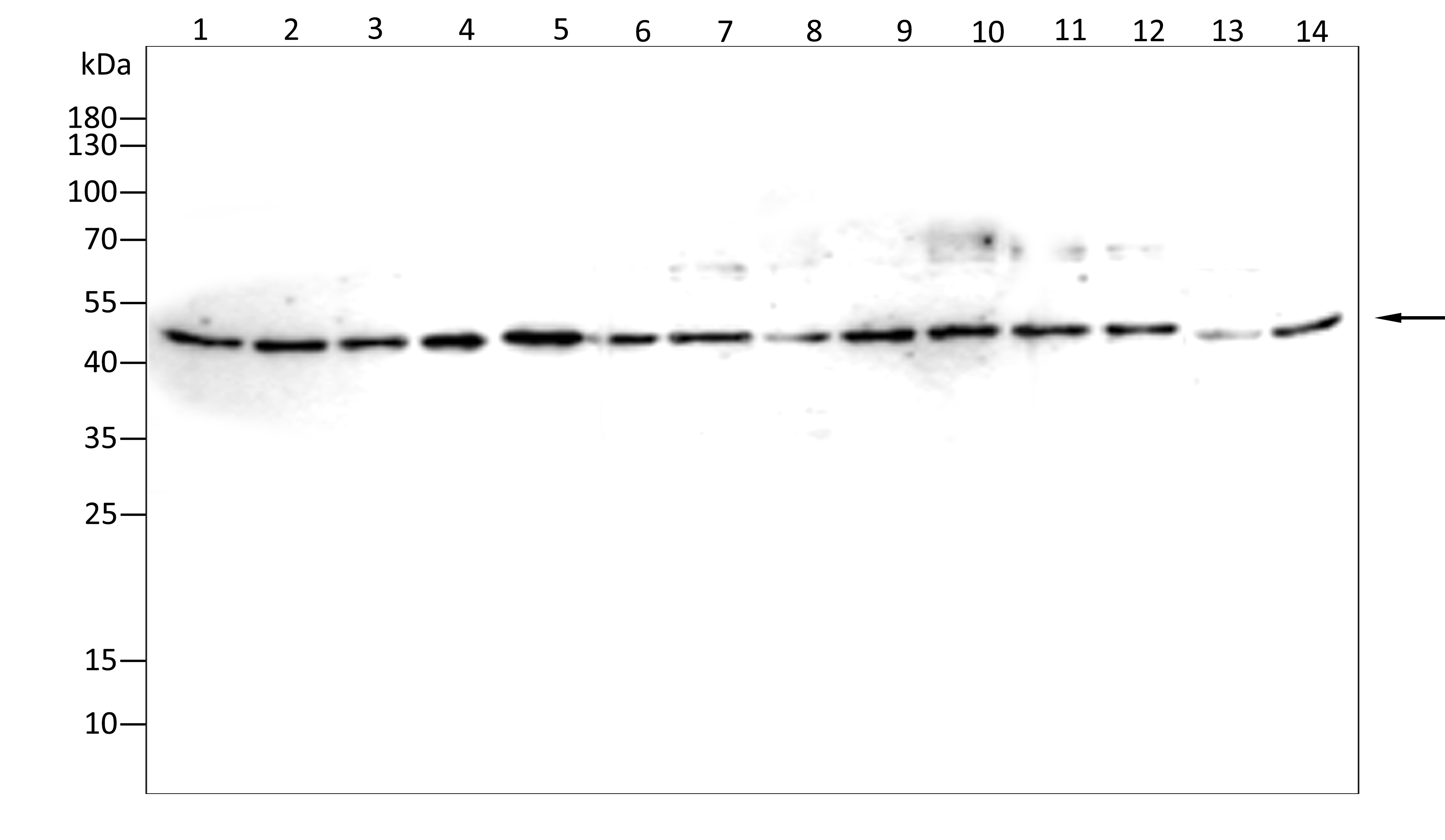
Western blot analysis using β-Actin antibody against Hela(1),Hepg2(2),A549(3),Huvec(4),Cos-7(5),P19(6),B16(7),C2C12(8),K562(9),Jurkat(10),Mfc(11),Nih3t3(12),T24(13),Raji(14) cell. 12% SDS-PAGE gel.Sample loading: 20μg /lane. Transfer the proteins onto a PVDF membrane (OM790003), and block it with TBST (OM750016) plus skimmed milk powder(OM750084) for one hour. Dilute the primary antibody with the antibody diluent (OM750012) at a ratio of 1:1000-1:2000, and incubate it overnight at 4°C. Wash the membrane three times with TBST (OM750016), 5 minutes each time. At room temperature, dilute the secondary antibody, Goat Anti-Rabbit IgG(H&L)-HRP (OM643487), at a ratio of 1:20000 and incubate for one hour. Wash the membrane three times with TBST (OM750016) again, 5 minutes each time. Use ECL (OM625701) for luminescence.staining time: 60S.WB
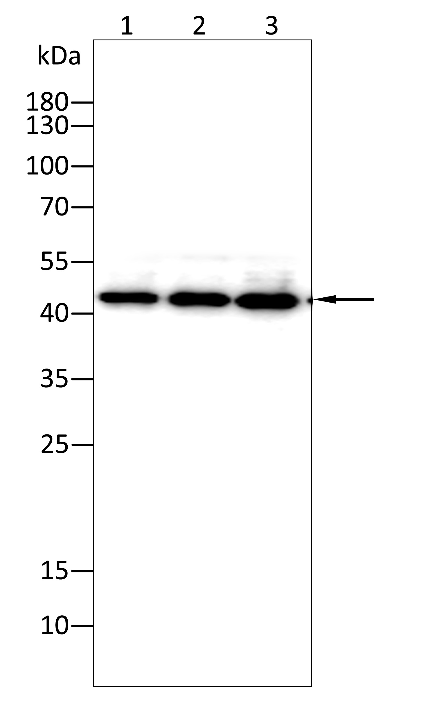
Western blot analysis using β-Actin antibody against Mouse brain(1),Mouse intestine(2),Mouse testes(3) tissue. 12% SDS-PAGE gel.Sample loading: 20μg /lane. Transfer the proteins onto a PVDF membrane (OM790003), and block it with TBST (OM750016) plus skimmed milk powder(OM750084) for one hour. Dilute the primary antibody with the antibody diluent (OM750012) at a ratio of 1:1000-1:2000, and incubate it overnight at 4°C. Wash the membrane three times with TBST (OM750016), 5 minutes each time. At room temperature, dilute the secondary antibody, Goat Anti-Rabbit IgG(H&L)-HRP (OM643487), at a ratio of 1:20000 and incubate for one hour. Wash the membrane three times with TBST (OM750016) again, 5 minutes each time. Use ECL (OM625701) for luminescence.staining time: 60S.ELISA
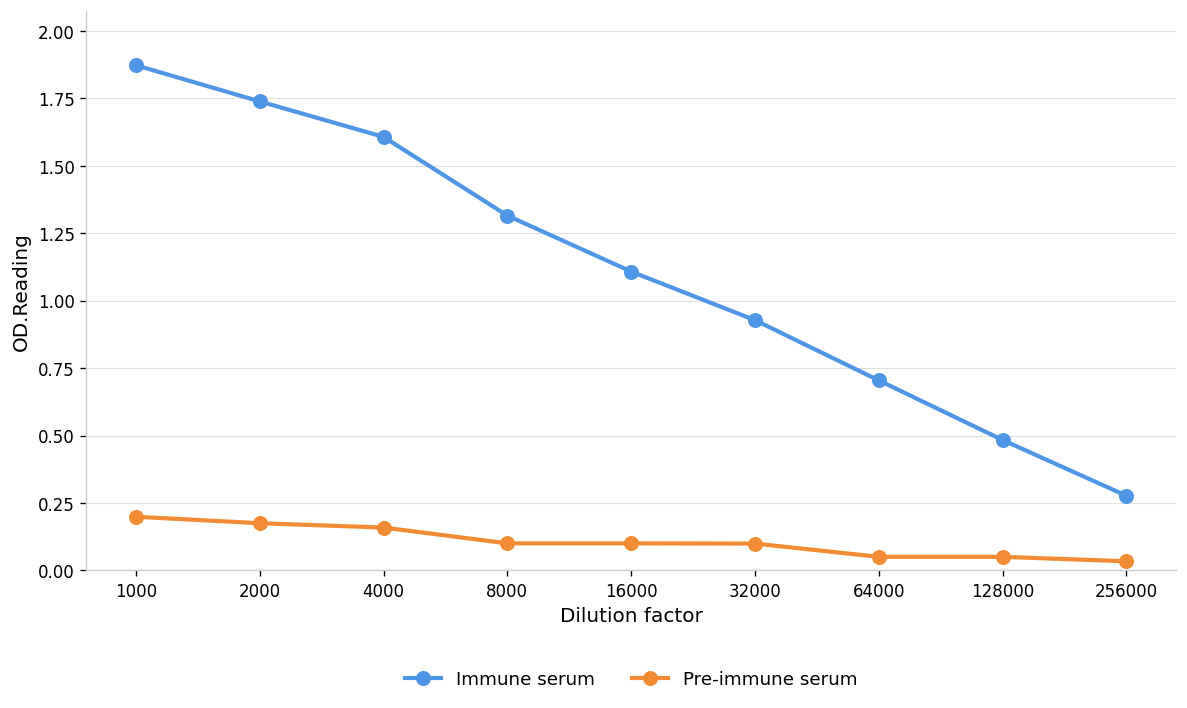
Coat the plate with immunizing antigen.Blue line: Immune serum; Orange line: Pre-immune serum.| Product Name | Anti-β-Actin antibody,pbs only |
|---|---|
| Antibody Type | Primary Antibodies |
| Immunogen | Polypeptide |
| Clonality | Polyclonal |
|---|---|
| Isotype | IgG |
| Host Species | Rabbit |
| Tested Applications | ELISAICC/IFWB |
| ICC/IF:1:100-1:500 WB:1:1000-1:2000 |
|
| Species Reactivity | HumanMouseRat |
| Concentration | 1mg/ml |
| Purification | Protein A |
| Gene Symbol | ACTB |
|---|---|
| Gene Synonyms | actb actin Actx Bact Beta-actin BKRNS BNS BRWS1 CSMH DDS1 E430023M04Rik PS1TP5BP1 THC8 XELAEV_18045052mg |
| Gene Full Name | actin beta |
| Gene Summary | This gene encodes one of six different actin proteins. Actins are highly conserved proteins that are involved in cell motility, structure, integrity, and intercellular signaling. The encoded protein is a major constituent of the contractile apparatus and one of the two nonmuscle cytoskeletal actins that are ubiquitously expressed. Mutations in this gene cause Baraitser-Winter syndrome 1, which is characterized by intellectual disability with a distinctive facial appearance in human patients. Numerous pseudogenes of this gene have been identified throughout the human genome. [provided by RefSeq, Aug 2017] |
| Alternative Names | A-X actin A-X actin-like protein Actin is a protein with a high degree of evolutionary conservation that undergoes polymerization to generate filaments which subsequently assemble into interconnected networks within the cytoplasm of cells. actin beta cytoplasmic Bact actin beta actin beta cytoskeletal actin beta-actin cytoplasmic 1 cytoplasmic beta-actin cytoplasmic protein cytoskeletal beta actin I(2)-actin melanoma X-actin PS1TP5-binding protein 1 |
| Molecular Weight(MW) | 42kDa |
| Cellular Localization | Nucleus,Cytoskeleton |
| SwissProt ID | P60709 |
|---|
ICC/IF
Immunofluorescence analysis of Hela cells using β-Actin antibody (green). Blue: DAPI fluorescent DNA dye. Red: Actin filaments have been labeled with Omnimabs® 594-Phalloidin.Cells are fixed in 4% paraformaldehyde at room temperature for 15 minutes. Then, they are permeabilized with a PBS (OM750003) solution containing 0.1% Triton X-100(OM750021) at room temperature for 15 minutes. Subsequently, the cells are blocked with 10% non - immune goat serum(OM760028) at room temperature for 1 hour.The cells are incubated overnight at 4°C with the primary antibody diluted 1:100 in PBS. The secondary antibody, Omnimabs® 488 Goat Ant-Rabbit IgG(H&L) (Green,OM643486), is diluted at a ratio of 1:400 and incubated with the cells for 1 hour.Nuclear DNA is labeled with DAPI (Blue,OM643160). F-actin is stained with Omnimabs® 594-Phalloidin (Red,OM750007) diluted 1:100 for 30 minutes.
WB
Western blot analysis using β-Actin antibody against Hela(1),Hepg2(2),A549(3),Huvec(4),Cos-7(5),P19(6),B16(7),C2C12(8),K562(9),Jurkat(10),Mfc(11),Nih3t3(12),T24(13),Raji(14) cell. 12% SDS-PAGE gel.Sample loading: 20μg /lane. Transfer the proteins onto a PVDF membrane (OM790003), and block it with TBST (OM750016) plus skimmed milk powder(OM750084) for one hour. Dilute the primary antibody with the antibody diluent (OM750012) at a ratio of 1:1000-1:2000, and incubate it overnight at 4°C. Wash the membrane three times with TBST (OM750016), 5 minutes each time. At room temperature, dilute the secondary antibody, Goat Anti-Rabbit IgG(H&L)-HRP (OM643487), at a ratio of 1:20000 and incubate for one hour. Wash the membrane three times with TBST (OM750016) again, 5 minutes each time. Use ECL (OM625701) for luminescence.staining time: 60S.
WB
Western blot analysis using β-Actin antibody against Mouse brain(1),Mouse intestine(2),Mouse testes(3) tissue. 12% SDS-PAGE gel.Sample loading: 20μg /lane. Transfer the proteins onto a PVDF membrane (OM790003), and block it with TBST (OM750016) plus skimmed milk powder(OM750084) for one hour. Dilute the primary antibody with the antibody diluent (OM750012) at a ratio of 1:1000-1:2000, and incubate it overnight at 4°C. Wash the membrane three times with TBST (OM750016), 5 minutes each time. At room temperature, dilute the secondary antibody, Goat Anti-Rabbit IgG(H&L)-HRP (OM643487), at a ratio of 1:20000 and incubate for one hour. Wash the membrane three times with TBST (OM750016) again, 5 minutes each time. Use ECL (OM625701) for luminescence.staining time: 60S.
ELISA
Coat the plate with immunizing antigen.Blue line: Immune serum; Orange line: Pre-immune serum.| Application Notes | ICC/IF:1:100-1:500 WB:1:1000-1:2000 |
|---|
| Form | Liquid |
|---|---|
| Storage Instructions | Shipped at 4°C. Store at +4°C short term (1-2 weeks). Store at -20°C long term. Avoid freeze / thaw cycle. |
| Storage Buffer | PBS ONLY |


