WB
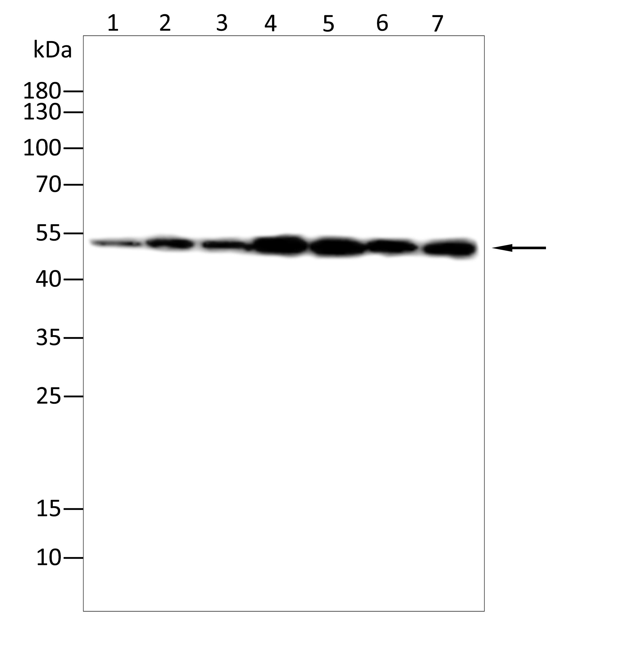
Western blot analysis using Vimentin mouse monoclonal antibody against Hela(1),Hepg2(2),A549(3),Huvec(4),Cos-7(5),P19(6),B16(7) cell lysate . 12% SDS-PAGE gel.Sample loading: 20μg /lane. Transfer the proteins onto a PVDF membrane (OM790003), and block it with TBST (OM750016) plus skimmed milk powder(OM750084) for one hour. Dilute the primary antibody with the antibody diluent (OM750012) at a ratio of 1:1000, and incubate it overnight at 4°C. Wash the membrane three times with TBST (OM750016), 5 minutes each time. At room temperature, dilute the secondary antibody, Goat Anti-Mouse IgG (H&L) - HRP(OM644366), at a ratio of 1:20000 and incubate for one hour. Wash the membrane three times with TBST (OM750016) again, 5 minutes each time. Use ECL (OM625701) for luminescence.staining time: 60SIHC
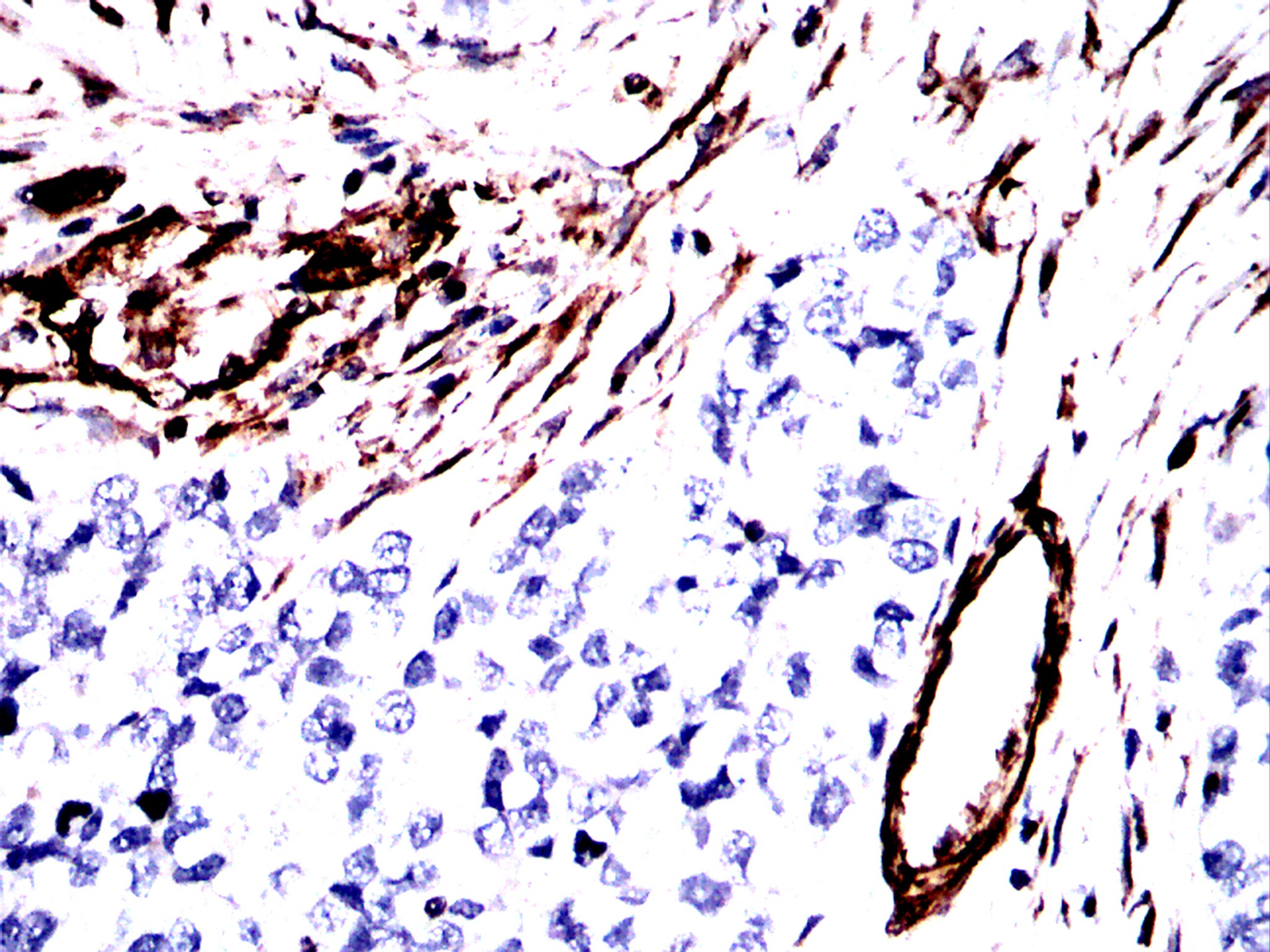
Immunohistochemical analysis of paraffin-embedded Human bladder cancer tissues using Vimentin antibody with DAB staining.Pre-treat the sections with heat-mediated antigen retrieval using sodium citrate buffer (pH 6.0) (OM750020) for 2 minutes. Wash the sections with ddH₂O and PBS (OM750003). Block the tissue with 10% non-immune goat serum(OM760028) at room temperature for 30 minutes. Incubate the tissue with the primary antibody diluted at a ratio of 1:1000 at 4°C overnight. At room temperature, dilute the secondary antibody, Goat Anti-Mouse IgG(H&L)-HRP (OM644366), at a ratio of 1:200 and incubate for one hour. Use DAB(OM760029)as the chromogenic agent. Counterstain the tissue with hematoxylin, and mount the tissue sections with neutral gum.IHC
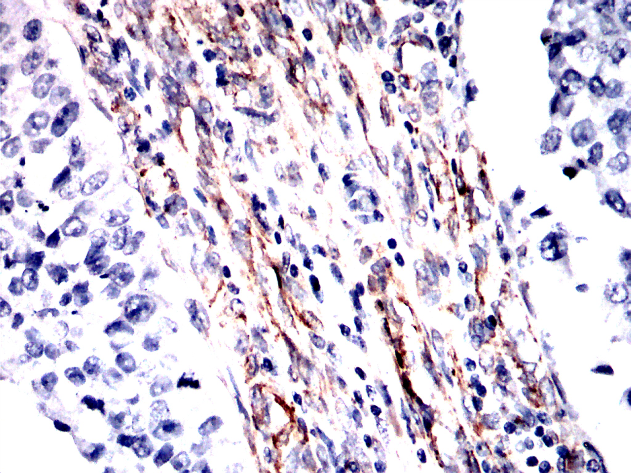
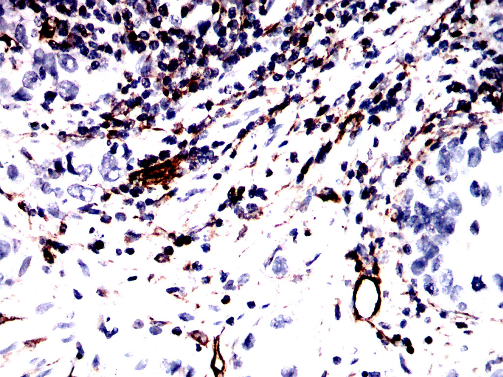
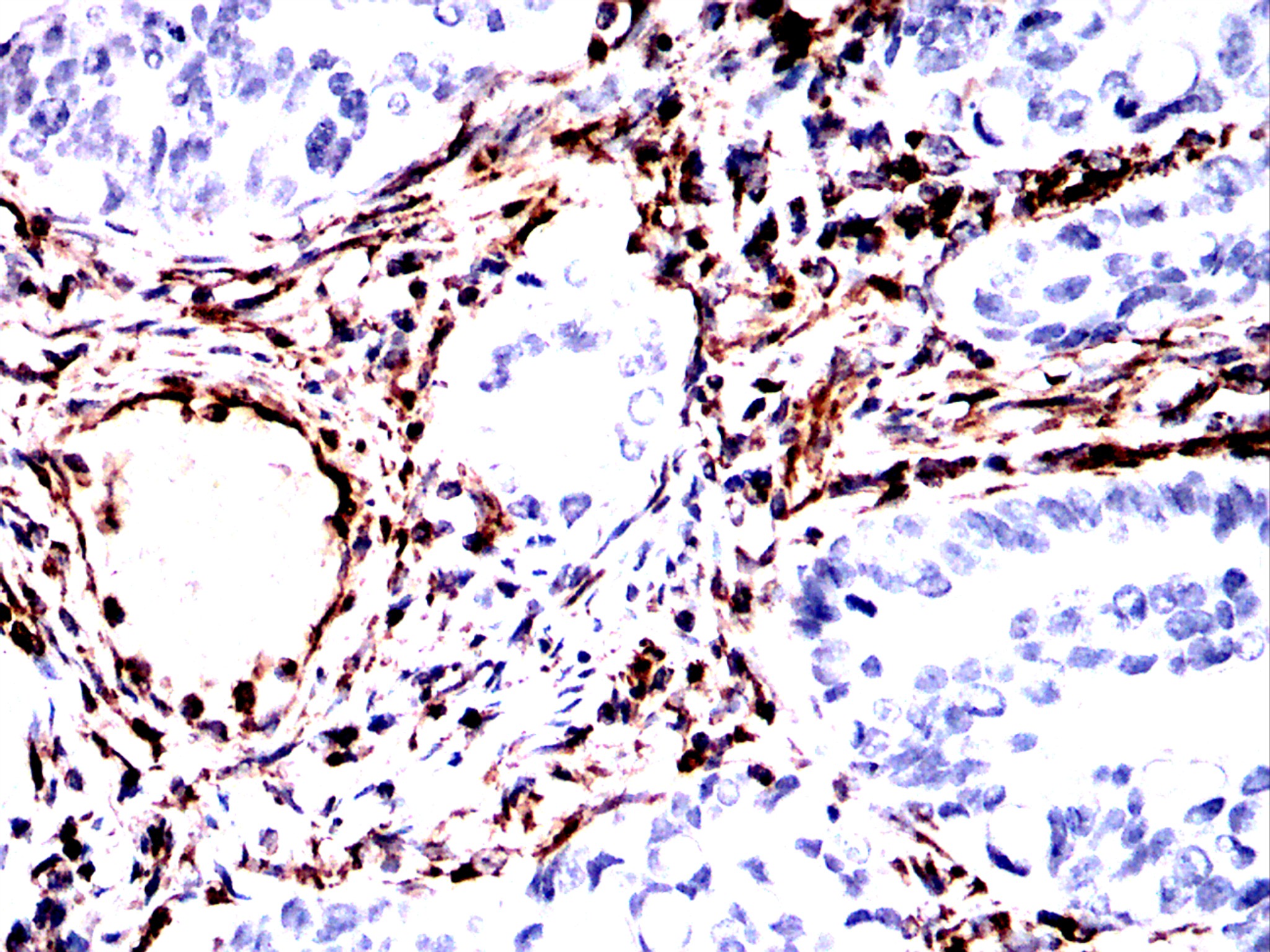
Immunohistochemical analysis of paraffin-embedded Human cervical cancer tissues using Vimentin antibody with DAB staining.Pre-treat the sections with heat-mediated antigen retrieval using sodium citrate buffer (pH 6.0) (OM750020) for 2 minutes. Wash the sections with ddH₂O and PBS (OM750003). Block the tissue with 10% non-immune goat serum(OM760028) at room temperature for 30 minutes. Incubate the tissue with the primary antibody diluted at a ratio of 1:1000 at 4°C overnight. At room temperature, dilute the secondary antibody, Goat Anti-Mouse IgG(H&L)-HRP (OM644366), at a ratio of 1:200 and incubate for one hour. Use DAB(OM760029)as the chromogenic agent. Counterstain the tissue with hematoxylin, and mount the tissue sections with neutral gum.IHC
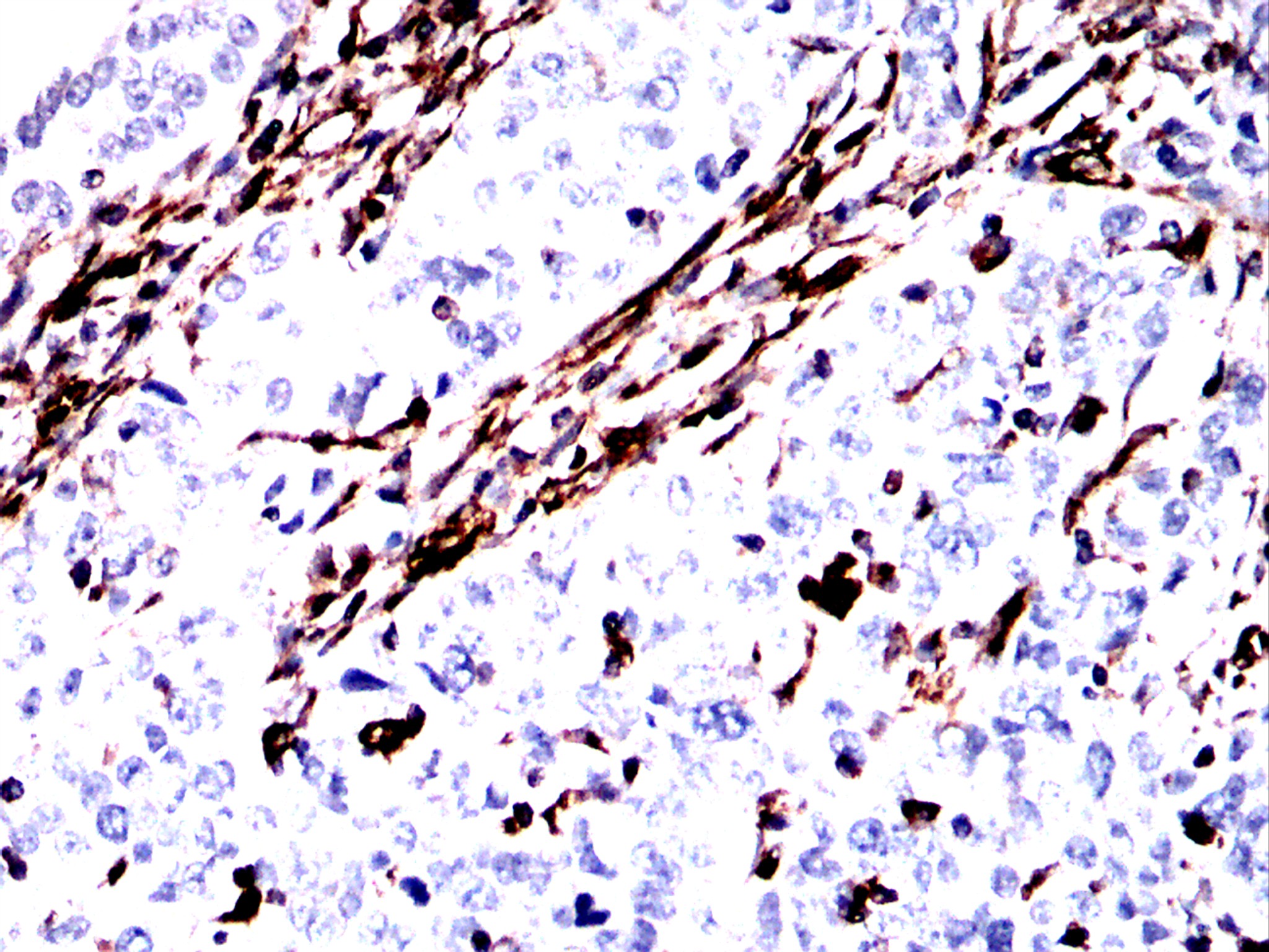
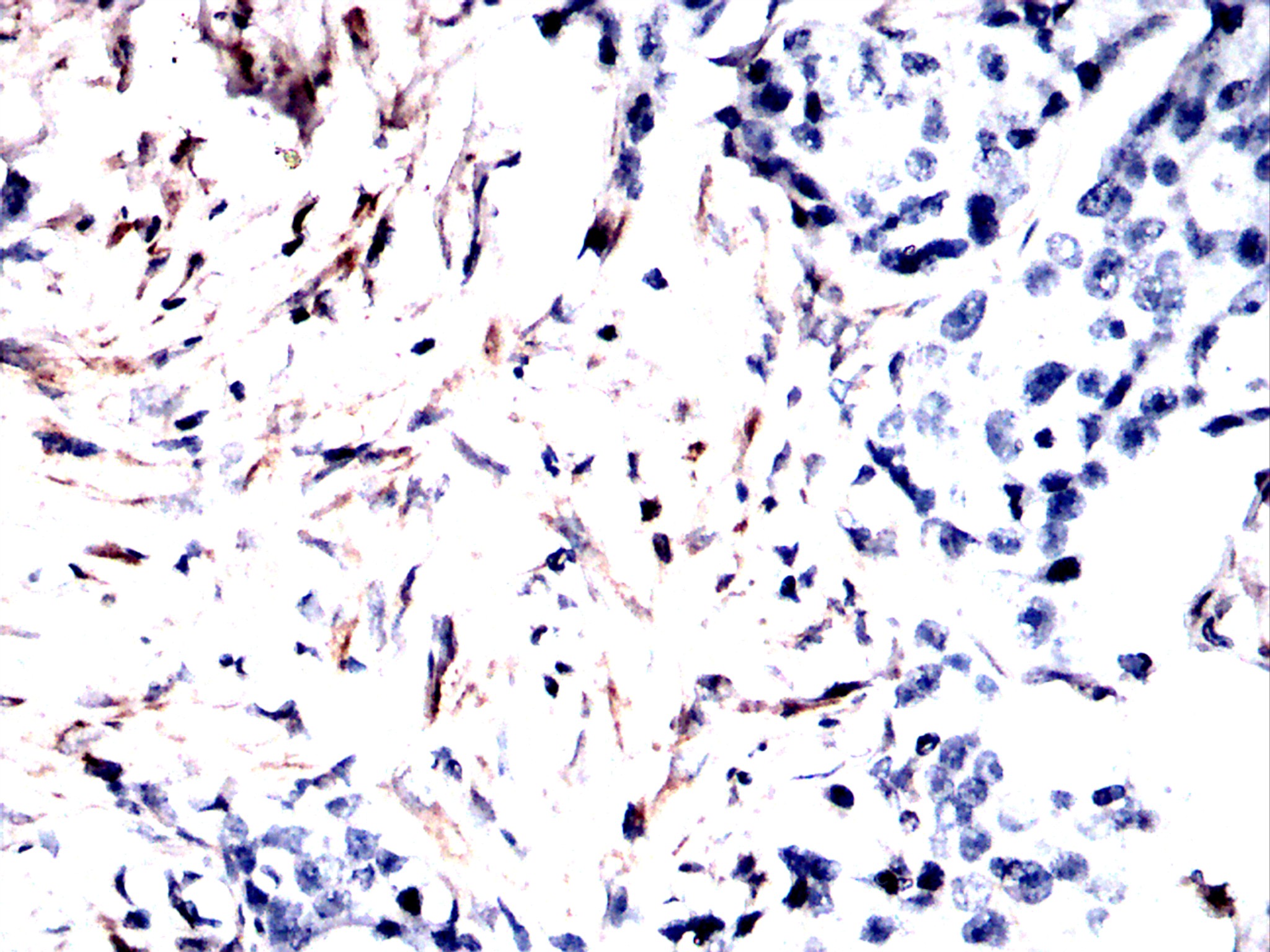
Immunohistochemical analysis of paraffin-embedded Human cervical cancer tissues using Vimentin antibody with DAB staining.Pre-treat the sections with heat-mediated antigen retrieval using sodium citrate buffer (pH 6.0) (OM750020) for 2 minutes. Wash the sections with ddH₂O and PBS (OM750003). Block the tissue with 10% non-immune goat serum(OM760028) at room temperature for 30 minutes. Incubate the tissue with the primary antibody diluted at a ratio of 1:1000 at 4°C overnight. At room temperature, dilute the secondary antibody, Goat Anti-Mouse IgG(H&L)-HRP (OM644366), at a ratio of 1:200 and incubate for one hour. Use DAB(OM760029)as the chromogenic agent. Counterstain the tissue with hematoxylin, and mount the tissue sections with neutral gum.IHC
Immunohistochemical analysis of paraffin-embedded Human ovarian cancer tissues using Vimentin antibody with DAB staining.Pre-treat the sections with heat-mediated antigen retrieval using sodium citrate buffer (pH 6.0) (OM750020) for 2 minutes. Wash the sections with ddH₂O and PBS (OM750003). Block the tissue with 10% non-immune goat serum(OM760028) at room temperature for 30 minutes. Incubate the tissue with the primary antibody diluted at a ratio of 1:1000 at 4°C overnight. At room temperature, dilute the secondary antibody, Goat Anti-Mouse IgG(H&L)-HRP (OM644366), at a ratio of 1:200 and incubate for one hour. Use DAB(OM760029)as the chromogenic agent. Counterstain the tissue with hematoxylin, and mount the tissue sections with neutral gum.IHC
Immunohistochemical analysis of paraffin-embedded Human prostate cancer tissues using Vimentin antibody with DAB staining.Pre-treat the sections with heat-mediated antigen retrieval using sodium citrate buffer (pH 6.0) (OM750020) for 2 minutes. Wash the sections with ddH₂O and PBS (OM750003). Block the tissue with 10% non-immune goat serum(OM760028) at room temperature for 30 minutes. Incubate the tissue with the primary antibody diluted at a ratio of 1:1000 at 4°C overnight. At room temperature, dilute the secondary antibody, Goat Anti-Mouse IgG(H&L)-HRP (OM644366), at a ratio of 1:200 and incubate for one hour. Use DAB(OM760029)as the chromogenic agent. Counterstain the tissue with hematoxylin, and mount the tissue sections with neutral gum.IHC
Immunohistochemical analysis of paraffin-embedded Human rectal cancer tissues using Vimentin antibody with DAB staining.Pre-treat the sections with heat-mediated antigen retrieval using sodium citrate buffer (pH 6.0) (OM750020) for 2 minutes. Wash the sections with ddH₂O and PBS (OM750003). Block the tissue with 10% non-immune goat serum(OM760028) at room temperature for 30 minutes. Incubate the tissue with the primary antibody diluted at a ratio of 1:1000 at 4°C overnight. At room temperature, dilute the secondary antibody, Goat Anti-Mouse IgG(H&L)-HRP (OM644366), at a ratio of 1:200 and incubate for one hour. Use DAB(OM760029)as the chromogenic agent. Counterstain the tissue with hematoxylin, and mount the tissue sections with neutral gum.ICC/IF
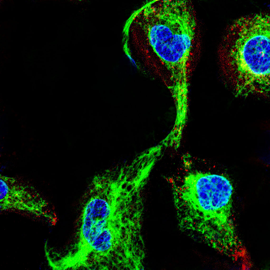
Immunofluorescence analysis of Hela cells using Vimentin antibody (green). Blue: DAPI fluorescent DNA dye. Red: Actin filaments have been labeled with Omnimabs® 594-Phalloidin.Cells are fixed in 4% paraformaldehyde at room temperature for 15 minutes. Then, they are permeabilized with a PBS (OM750003) solution containing 0.1% Triton X-100(OM750021) at room temperature for 15 minutes. Subsequently, the cells are blocked with 10% non - immune goat serum(OM760028) at room temperature for 1 hour.The cells are incubated overnight at 4°C with the primary antibody diluted 1:100 in PBS. The secondary antibody, Omnimabs® 488 Goat Ant-Mouse IgG(H&L) (Green,OM643485), is diluted at a ratio of 1:400 and incubated with the cells for 1 hour.Nuclear DNA is labeled with DAPI (Blue,OM643160). F-actin is stained with Omnimabs® 594-Phalloidin (Red,OM750007) diluted 1:100 for 30 minutes.IF-P
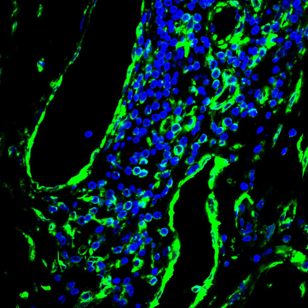
Immunohistochemical analysis of paraffin-embedded Human stomach cancer tissues using Vimentin antibody with DAB staining.Pre-treat the sections with heat-mediated antigen retrieval using sodium citrate buffer (pH 6.0) (OM750020) for 2 minutes. Wash the sections with ddH₂O and PBS (OM750003). Block the tissue with 10% non-immune goat serum(OM760028) at room temperature for 30 minutes. Incubate the tissue with the primary antibody diluted at a ratio of 1:200 at 4°C overnight. At room temperature, dilute the secondary antibody, Goat Anti-Mouse IgG(H&L)-HRP (OM644366), at a ratio of 1:200 and incubate for one hour. Tyramine labeled with 488 (Green,OM642679) was used as chromogenic agent, and DAPI(Blue,OM642679) was used for double dyeing, and the anti-fluorescence attenuation tablets were sealed.ELISA
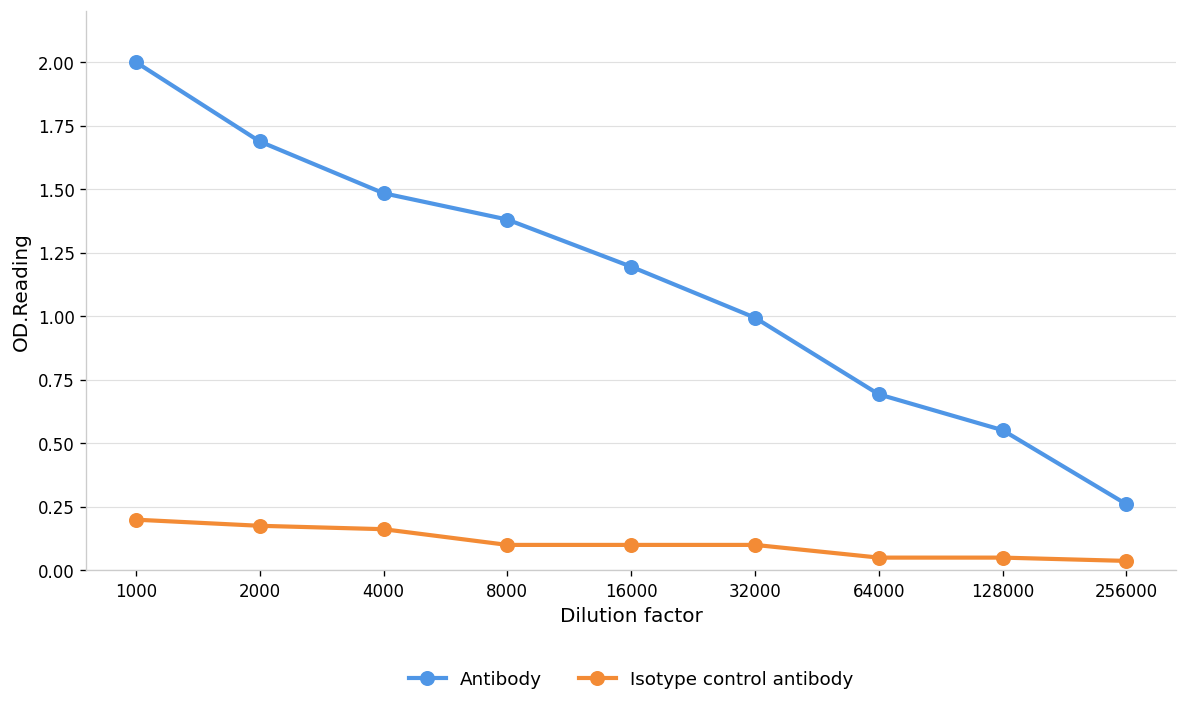
Coat the plate with immunizing antigen.Blue line: Antibody; Orange line: Isotype control antibody| Product Name | Anti-Vimentin antibody,pbs only |
|---|---|
| Antibody Type | Primary Antibodies |
| Immunogen | Recombinant protein |
| Clonality | Monoclonal |
|---|---|
| Isotype | IgG |
| Host Species | Mouse |
| Tested Applications | ELISAICC/IFIF-PIHCWB |
| WB:1:1000 IHC:1:1000 ICC/IF:1:100-1:500 IF-P:1:200-1:2000 |
|
| Species Reactivity | HumanMouseRat |
| Concentration | 1mg/ml |
| Purification | Affinity purified |
| Gene Symbol | VIM |
|---|---|
| Gene Synonyms | cb28 |
| Gene Full Name | vimentin |
| Gene Summary | This gene encodes a type III intermediate filament protein. Intermediate filaments, along with microtubules and actin microfilaments, make up the cytoskeleton. The encoded protein is responsible for maintaining cell shape and integrity of the cytoplasm, and stabilizing cytoskeletal interactions. This protein is involved in neuritogenesis and cholesterol transport and functions as an organizer of a number of other critical proteins involved in cell attachment, migration, and signaling. Bacterial and viral pathogens have been shown to attach to this protein on the host cell surface. Mutations in this gene are associated with congenital cataracts in human patients. [provided by RefSeq, Aug 2017] |
| Alternative Names | epididymis secretory sperm binding protein FLJ36605 Hel113 intermediate filament intermediate filament protein RP11-124N14.1 VIM VIME vimentin vimentin C-terminus vimentin protein |
| Molecular Weight(MW) | 54kDA |
| Function | Vimentin (57 kDa) is the most ubiquituos intermediate filament protein and the first to be expressed during cell differentiation. All primitive cell types express vimentin but in most non-mesenchymal cells it is replaced by other intermediate filament proteins during differentiation. Vimentin is expressed in a wide variety of mesenchymal cell types: fibroblasts, endothelial cells etc., and in a number of other cell types derived from mesoderm, e.g., mesothelium and ovarian granulosa cells. Vimentin is present in many different neoplasms but is particulary expressed in those originated from mesenchymal cells. Sarcomas e.g., fibrosarcoma, malignt fibrous histiocytoma, angiosarcoma, and leio- and rhabdomyosarcoma, as well as lymphomas, malignant melanoma and schwannoma, are virtually always vimentin positive. Mesoderm derived carcinomas like renal cell carcinoma, adrenal cortical carcinoma and adenocarcinomas from endometrium and ovary usually express vimentin. Also thyroid carcinomas are vimentin positive. Any low differentiated or sarcomatoid carcinoma may express some vimentin. Vimentin is frequently included in the so-called primary panel (together with CD45, cytokeratin, and S-100 protein): Intense staining reaction for vimentin without coexpression of other intermediate filament proteins is strongly suggestive of a mesenchymal tumour or a malignant melanoma. However, in biopsies representing only a sarcomatoid part of renal cell carcinoma a.o. a strong positivity for vimentin without cytokeratin expression may be seen. Tumours like lymphomas and seminomas have the same intermediate filament profile, but the vimentin expression is usually weaker. |
| Cellular Localization | Cell membrane Cytoplasm Cytoskeleton Intermediate filament Membrane Nucleus |
| SwissProt ID | P08670 |
|---|
WB
Western blot analysis using Vimentin mouse monoclonal antibody against Hela(1),Hepg2(2),A549(3),Huvec(4),Cos-7(5),P19(6),B16(7) cell lysate . 12% SDS-PAGE gel.Sample loading: 20μg /lane. Transfer the proteins onto a PVDF membrane (OM790003), and block it with TBST (OM750016) plus skimmed milk powder(OM750084) for one hour. Dilute the primary antibody with the antibody diluent (OM750012) at a ratio of 1:1000, and incubate it overnight at 4°C. Wash the membrane three times with TBST (OM750016), 5 minutes each time. At room temperature, dilute the secondary antibody, Goat Anti-Mouse IgG (H&L) - HRP(OM644366), at a ratio of 1:20000 and incubate for one hour. Wash the membrane three times with TBST (OM750016) again, 5 minutes each time. Use ECL (OM625701) for luminescence.staining time: 60S
IHC
Immunohistochemical analysis of paraffin-embedded Human bladder cancer tissues using Vimentin antibody with DAB staining.Pre-treat the sections with heat-mediated antigen retrieval using sodium citrate buffer (pH 6.0) (OM750020) for 2 minutes. Wash the sections with ddH₂O and PBS (OM750003). Block the tissue with 10% non-immune goat serum(OM760028) at room temperature for 30 minutes. Incubate the tissue with the primary antibody diluted at a ratio of 1:1000 at 4°C overnight. At room temperature, dilute the secondary antibody, Goat Anti-Mouse IgG(H&L)-HRP (OM644366), at a ratio of 1:200 and incubate for one hour. Use DAB(OM760029)as the chromogenic agent. Counterstain the tissue with hematoxylin, and mount the tissue sections with neutral gum.
IHC
Immunohistochemical analysis of paraffin-embedded Human cervical cancer tissues using Vimentin antibody with DAB staining.Pre-treat the sections with heat-mediated antigen retrieval using sodium citrate buffer (pH 6.0) (OM750020) for 2 minutes. Wash the sections with ddH₂O and PBS (OM750003). Block the tissue with 10% non-immune goat serum(OM760028) at room temperature for 30 minutes. Incubate the tissue with the primary antibody diluted at a ratio of 1:1000 at 4°C overnight. At room temperature, dilute the secondary antibody, Goat Anti-Mouse IgG(H&L)-HRP (OM644366), at a ratio of 1:200 and incubate for one hour. Use DAB(OM760029)as the chromogenic agent. Counterstain the tissue with hematoxylin, and mount the tissue sections with neutral gum.
IHC
Immunohistochemical analysis of paraffin-embedded Human cervical cancer tissues using Vimentin antibody with DAB staining.Pre-treat the sections with heat-mediated antigen retrieval using sodium citrate buffer (pH 6.0) (OM750020) for 2 minutes. Wash the sections with ddH₂O and PBS (OM750003). Block the tissue with 10% non-immune goat serum(OM760028) at room temperature for 30 minutes. Incubate the tissue with the primary antibody diluted at a ratio of 1:1000 at 4°C overnight. At room temperature, dilute the secondary antibody, Goat Anti-Mouse IgG(H&L)-HRP (OM644366), at a ratio of 1:200 and incubate for one hour. Use DAB(OM760029)as the chromogenic agent. Counterstain the tissue with hematoxylin, and mount the tissue sections with neutral gum.
IHC
Immunohistochemical analysis of paraffin-embedded Human ovarian cancer tissues using Vimentin antibody with DAB staining.Pre-treat the sections with heat-mediated antigen retrieval using sodium citrate buffer (pH 6.0) (OM750020) for 2 minutes. Wash the sections with ddH₂O and PBS (OM750003). Block the tissue with 10% non-immune goat serum(OM760028) at room temperature for 30 minutes. Incubate the tissue with the primary antibody diluted at a ratio of 1:1000 at 4°C overnight. At room temperature, dilute the secondary antibody, Goat Anti-Mouse IgG(H&L)-HRP (OM644366), at a ratio of 1:200 and incubate for one hour. Use DAB(OM760029)as the chromogenic agent. Counterstain the tissue with hematoxylin, and mount the tissue sections with neutral gum.
IHC
Immunohistochemical analysis of paraffin-embedded Human prostate cancer tissues using Vimentin antibody with DAB staining.Pre-treat the sections with heat-mediated antigen retrieval using sodium citrate buffer (pH 6.0) (OM750020) for 2 minutes. Wash the sections with ddH₂O and PBS (OM750003). Block the tissue with 10% non-immune goat serum(OM760028) at room temperature for 30 minutes. Incubate the tissue with the primary antibody diluted at a ratio of 1:1000 at 4°C overnight. At room temperature, dilute the secondary antibody, Goat Anti-Mouse IgG(H&L)-HRP (OM644366), at a ratio of 1:200 and incubate for one hour. Use DAB(OM760029)as the chromogenic agent. Counterstain the tissue with hematoxylin, and mount the tissue sections with neutral gum.
IHC
Immunohistochemical analysis of paraffin-embedded Human rectal cancer tissues using Vimentin antibody with DAB staining.Pre-treat the sections with heat-mediated antigen retrieval using sodium citrate buffer (pH 6.0) (OM750020) for 2 minutes. Wash the sections with ddH₂O and PBS (OM750003). Block the tissue with 10% non-immune goat serum(OM760028) at room temperature for 30 minutes. Incubate the tissue with the primary antibody diluted at a ratio of 1:1000 at 4°C overnight. At room temperature, dilute the secondary antibody, Goat Anti-Mouse IgG(H&L)-HRP (OM644366), at a ratio of 1:200 and incubate for one hour. Use DAB(OM760029)as the chromogenic agent. Counterstain the tissue with hematoxylin, and mount the tissue sections with neutral gum.
ICC/IF
Immunofluorescence analysis of Hela cells using Vimentin antibody (green). Blue: DAPI fluorescent DNA dye. Red: Actin filaments have been labeled with Omnimabs® 594-Phalloidin.Cells are fixed in 4% paraformaldehyde at room temperature for 15 minutes. Then, they are permeabilized with a PBS (OM750003) solution containing 0.1% Triton X-100(OM750021) at room temperature for 15 minutes. Subsequently, the cells are blocked with 10% non - immune goat serum(OM760028) at room temperature for 1 hour.The cells are incubated overnight at 4°C with the primary antibody diluted 1:100 in PBS. The secondary antibody, Omnimabs® 488 Goat Ant-Mouse IgG(H&L) (Green,OM643485), is diluted at a ratio of 1:400 and incubated with the cells for 1 hour.Nuclear DNA is labeled with DAPI (Blue,OM643160). F-actin is stained with Omnimabs® 594-Phalloidin (Red,OM750007) diluted 1:100 for 30 minutes.
IF-P
Immunohistochemical analysis of paraffin-embedded Human stomach cancer tissues using Vimentin antibody with DAB staining.Pre-treat the sections with heat-mediated antigen retrieval using sodium citrate buffer (pH 6.0) (OM750020) for 2 minutes. Wash the sections with ddH₂O and PBS (OM750003). Block the tissue with 10% non-immune goat serum(OM760028) at room temperature for 30 minutes. Incubate the tissue with the primary antibody diluted at a ratio of 1:200 at 4°C overnight. At room temperature, dilute the secondary antibody, Goat Anti-Mouse IgG(H&L)-HRP (OM644366), at a ratio of 1:200 and incubate for one hour. Tyramine labeled with 488 (Green,OM642679) was used as chromogenic agent, and DAPI(Blue,OM642679) was used for double dyeing, and the anti-fluorescence attenuation tablets were sealed.
ELISA
Coat the plate with immunizing antigen.Blue line: Antibody; Orange line: Isotype control antibody| Application Notes | WB:1:1000 IHC:1:1000 ICC/IF:1:100-1:500 IF-P:1:200-1:2000 |
|---|
| Form | Liquid |
|---|---|
| Storage Instructions | Shipped at 4°C. Store at +4°C short term (1-2 weeks). Store at -20°C long term. Avoid freeze / thaw cycle. |
| Storage Buffer | PBS ONLY |


