WB
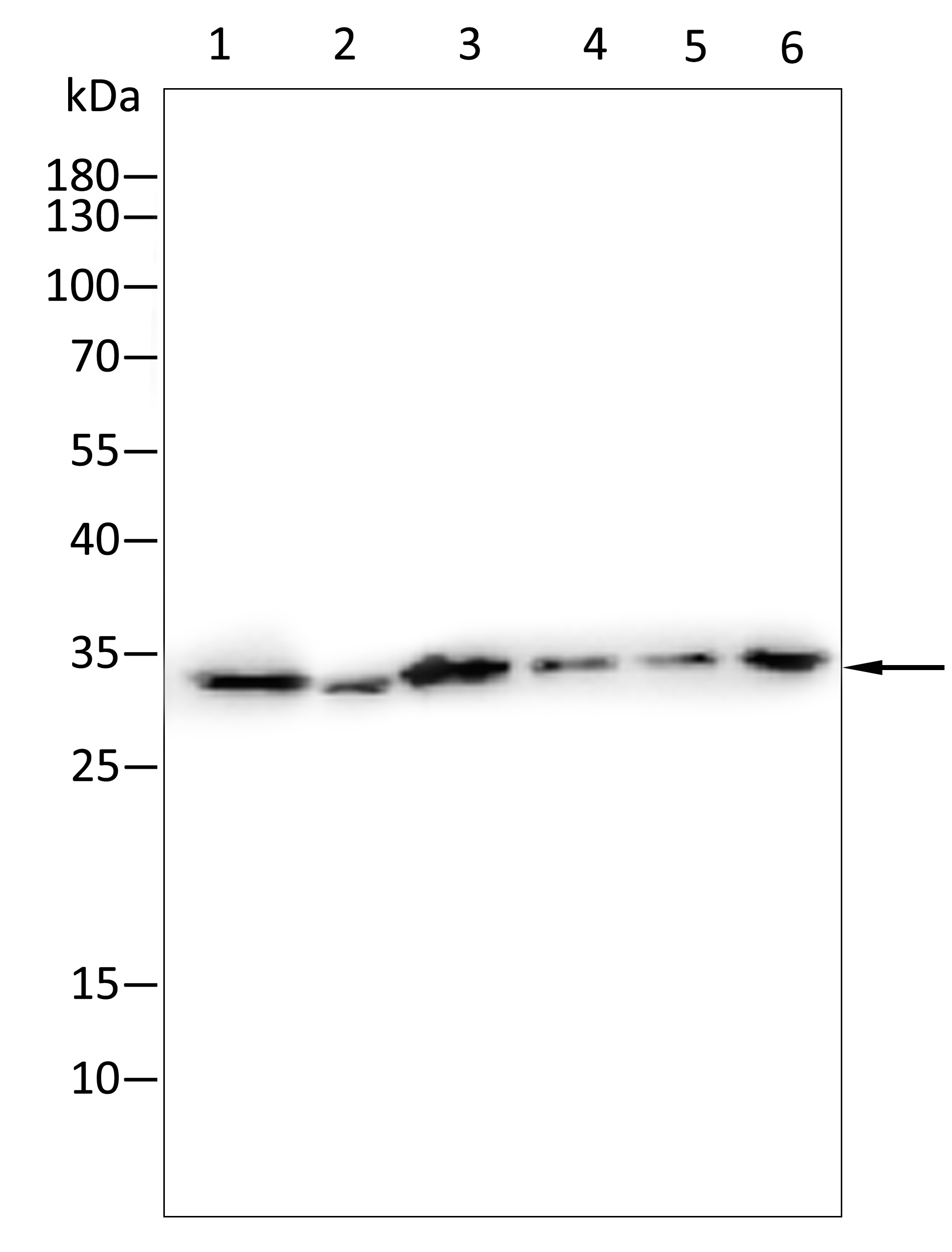
Western blot analysis using CDK5 mouse monoclonal antibody against Hela(1),Hepg2(2),A549(3),Huvec(4),Cos-7(5),P19(6) cell lysate . 12% SDS-PAGE gel.Sample loading: 20μg /lane. Transfer the proteins onto a PVDF membrane (OM790003), and block it with TBST (OM750016) plus skimmed milk powder(OM750084) for one hour. Dilute the primary antibody with the antibody diluent (OM750012) at a ratio of 1:1000, and incubate it overnight at 4°C. Wash the membrane three times with TBST (OM750016), 5 minutes each time. At room temperature, dilute the secondary antibody, Goat Anti-Mouse IgG (H&L) - HRP(OM644366), at a ratio of 1:20000 and incubate for one hour. Wash the membrane three times with TBST (OM750016) again, 5 minutes each time. Use ECL (OM625701) for luminescence.staining time: 60SIHC
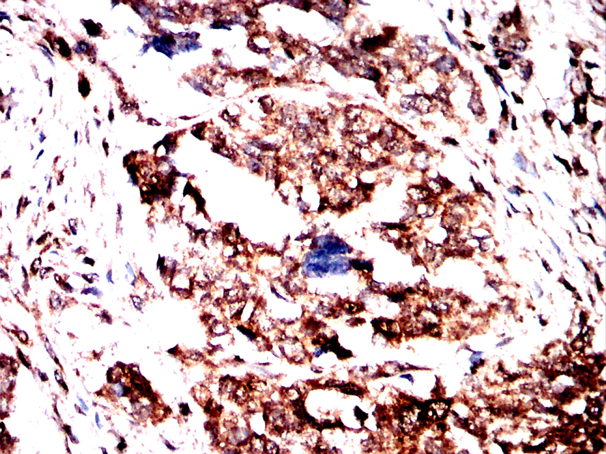
Immunohistochemical analysis of paraffin-embedded breast cancer tissues using CDK5 antibody with DAB staining.Pre-treat the sections with heat-mediated antigen retrieval using sodium citrate buffer (pH 6.0) (OM750020) for 2 minutes. Wash the sections with ddH₂O and PBS (OM750003). Block the tissue with 10% non-immune goat serum(OM760028) at room temperature for 30 minutes. Incubate the tissue with the primary antibody diluted at a ratio of 1:1000 at 4°C overnight. At room temperature, dilute the secondary antibody, Goat Anti-Mouse IgG(H&L)-HRP (OM644366), at a ratio of 1:200 and incubate for one hour. Use DAB(OM760029)as the chromogenic agent. Counterstain the tissue with hematoxylin, and mount the tissue sections with neutral gum.IHC
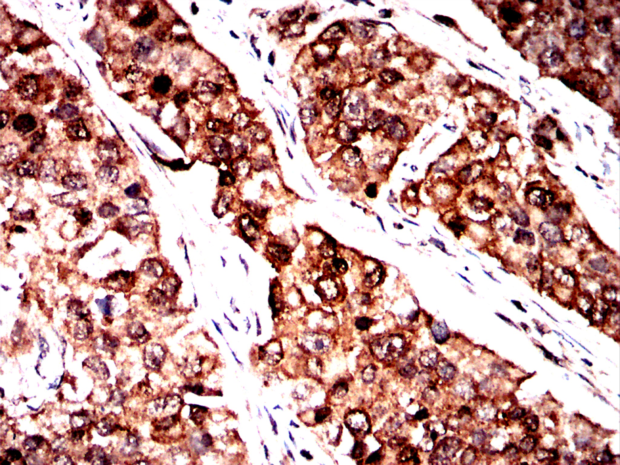
Immunohistochemical analysis of paraffin-embedded cervical cancer tissues using CDK5 antibody with DAB staining.Pre-treat the sections with heat-mediated antigen retrieval using sodium citrate buffer (pH 6.0) (OM750020) for 2 minutes. Wash the sections with ddH₂O and PBS (OM750003). Block the tissue with 10% non-immune goat serum(OM760028) at room temperature for 30 minutes. Incubate the tissue with the primary antibody diluted at a ratio of 1:1000 at 4°C overnight. At room temperature, dilute the secondary antibody, Goat Anti-Mouse IgG(H&L)-HRP (OM644366), at a ratio of 1:200 and incubate for one hour. Use DAB(OM760029)as the chromogenic agent. Counterstain the tissue with hematoxylin, and mount the tissue sections with neutral gum.IHC
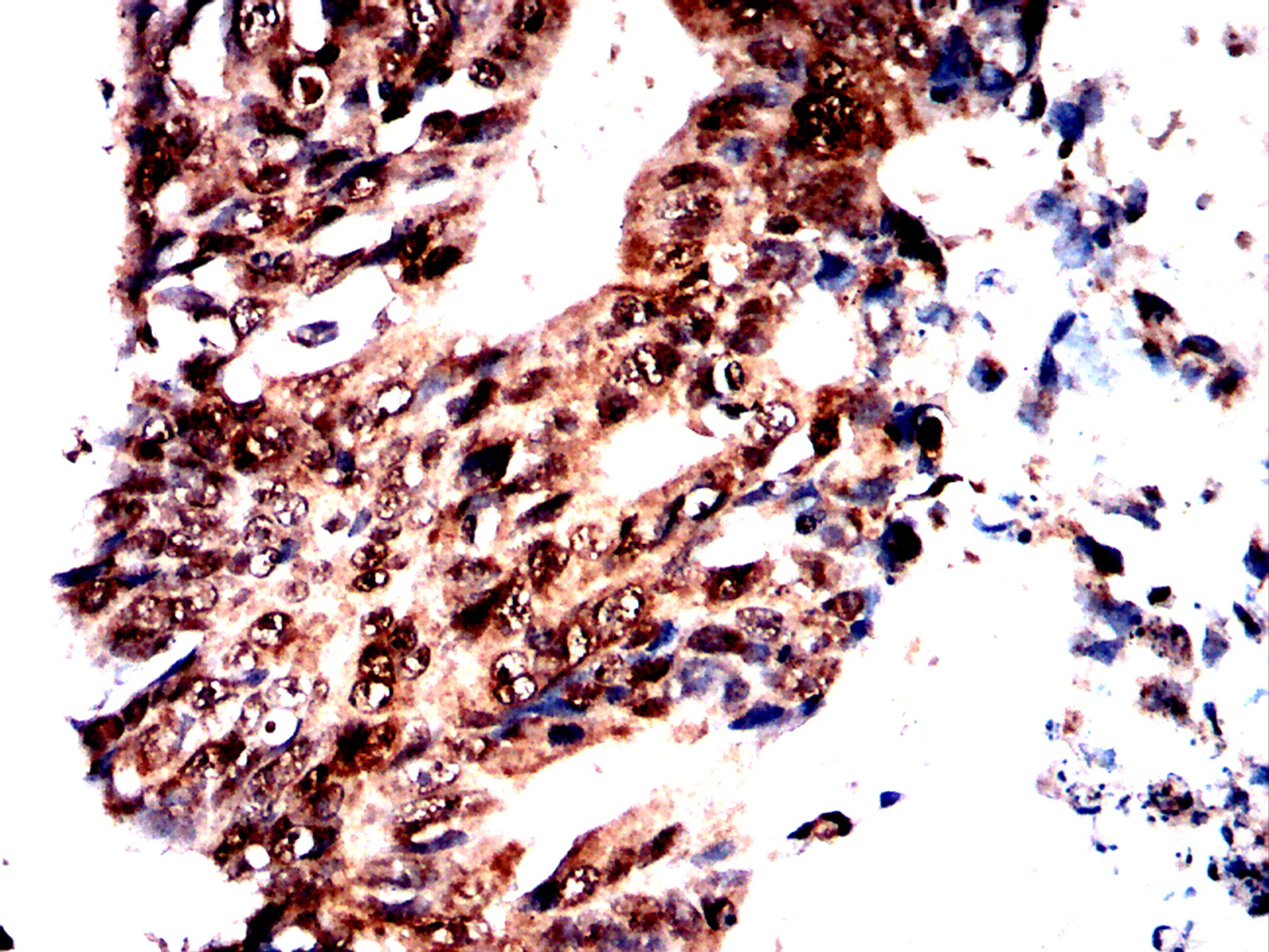
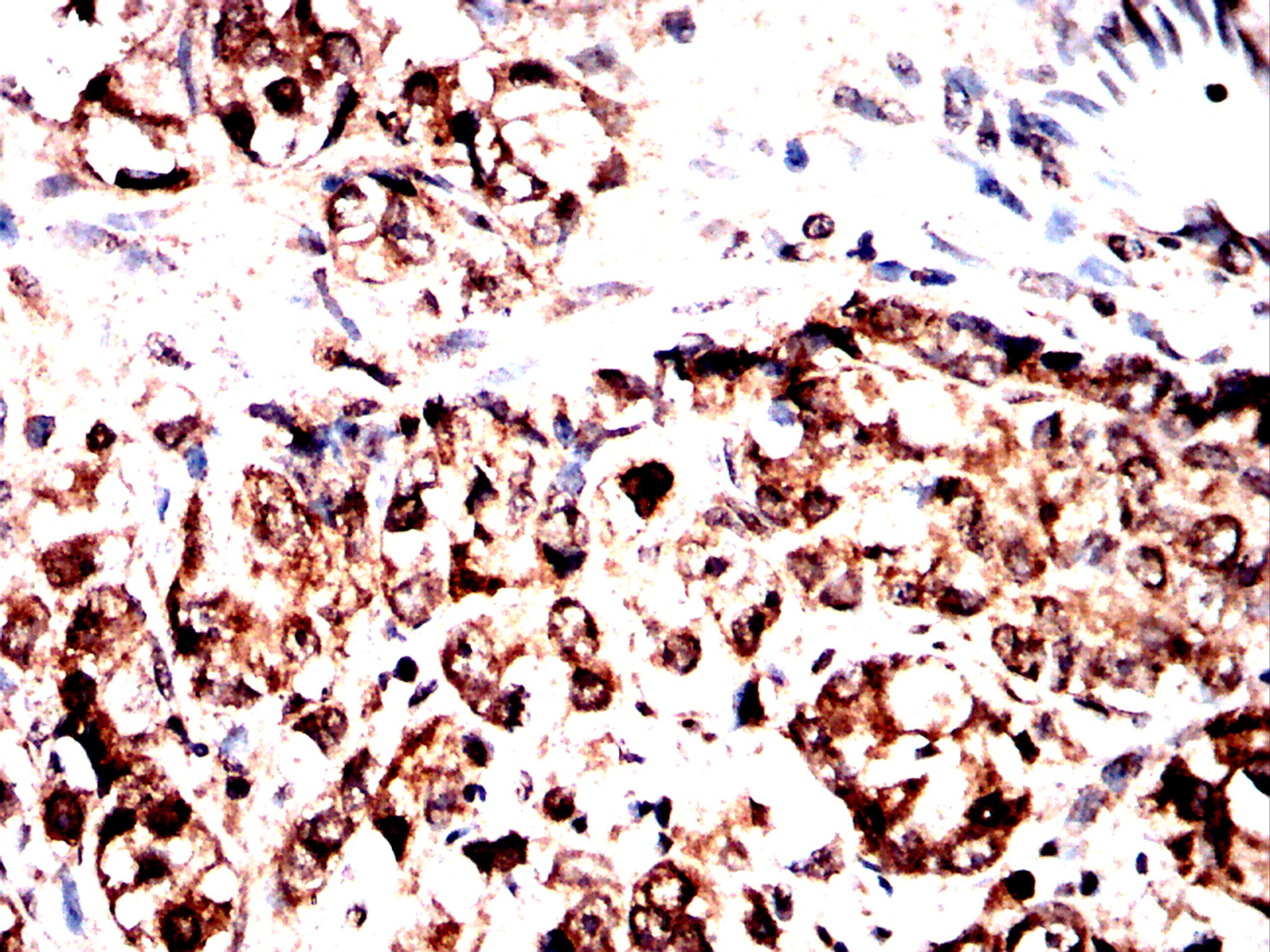
Immunohistochemical analysis of paraffin-embedded colon cancer tissues using CDK5 antibody with DAB staining.Pre-treat the sections with heat-mediated antigen retrieval using sodium citrate buffer (pH 6.0) (OM750020) for 2 minutes. Wash the sections with ddH₂O and PBS (OM750003). Block the tissue with 10% non-immune goat serum(OM760028) at room temperature for 30 minutes. Incubate the tissue with the primary antibody diluted at a ratio of 1:1000 at 4°C overnight. At room temperature, dilute the secondary antibody, Goat Anti-Mouse IgG(H&L)-HRP (OM644366), at a ratio of 1:200 and incubate for one hour. Use DAB(OM760029)as the chromogenic agent. Counterstain the tissue with hematoxylin, and mount the tissue sections with neutral gum.IHC
Immunohistochemical analysis of paraffin-embedded prostate cancer tissues using CDK5 antibody with DAB staining.Pre-treat the sections with heat-mediated antigen retrieval using sodium citrate buffer (pH 6.0) (OM750020) for 2 minutes. Wash the sections with ddH₂O and PBS (OM750003). Block the tissue with 10% non-immune goat serum(OM760028) at room temperature for 30 minutes. Incubate the tissue with the primary antibody diluted at a ratio of 1:1000 at 4°C overnight. At room temperature, dilute the secondary antibody, Goat Anti-Mouse IgG(H&L)-HRP (OM644366), at a ratio of 1:200 and incubate for one hour. Use DAB(OM760029)as the chromogenic agent. Counterstain the tissue with hematoxylin, and mount the tissue sections with neutral gum.ICC/IF
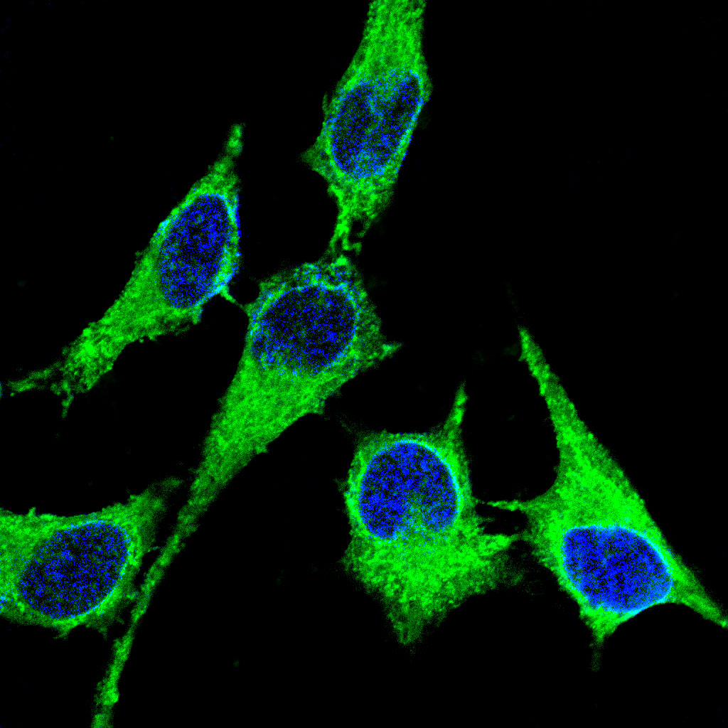
Immunofluorescence analysis of Hela cells using CDK5 antibody (green). Blue: DAPI fluorescent DNA dye.Cells are fixed in 4% paraformaldehyde at room temperature for 15 minutes. Then, they are permeabilized with a PBS (OM750003) solution containing 0.1% Triton X-100(OM750021) at room temperature for 15 minutes. Subsequently, the cells are blocked with 10% non - immune goat serum(OM760028) at room temperature for 1 hour.The cells are incubated overnight at 4°C with the primary antibody diluted 1:100 in PBS. The secondary antibody, Omnimabs® 488 Goat Ant-Mouse IgG(H&L) (Green,OM643485), is diluted at a ratio of 1:400 and incubated with the cells for 1 hour.Nuclear DNA is labeled with DAPI (Blue,OM643160).IF-P
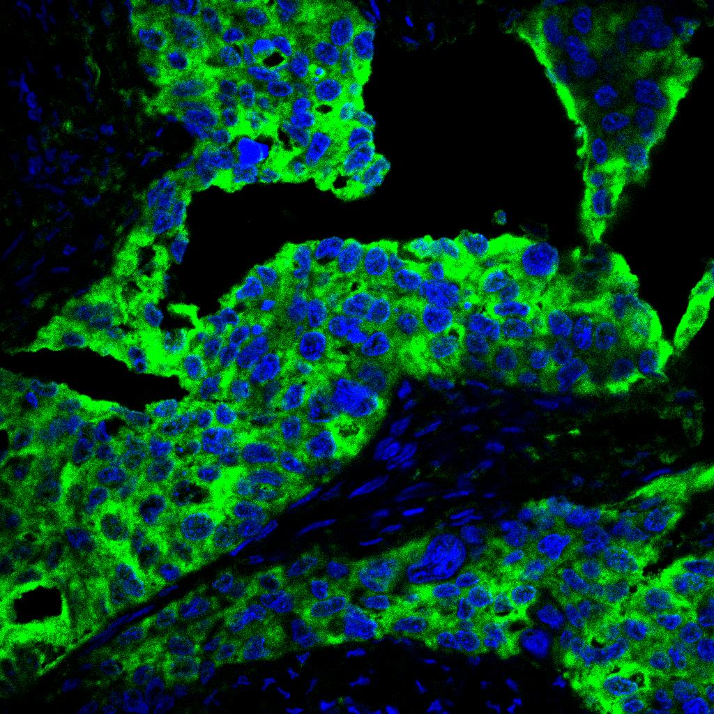
Immunohistochemical analysis of paraffin-embedded human cervical cancer tissues using CDK5 antibody with DAB staining.Pre-treat the sections with heat-mediated antigen retrieval using sodium citrate buffer (pH 6.0) (OM750020) for 2 minutes. Wash the sections with ddH₂O and PBS (OM750003). Block the tissue with 10% non-immune goat serum(OM760028) at room temperature for 30 minutes. Incubate the tissue with the primary antibody diluted at a ratio of 1:200 at 4°C overnight. At room temperature, dilute the secondary antibody, Goat Anti-Mouse IgG(H&L)-HRP (OM644366), at a ratio of 1:200 and incubate for one hour. Tyramine labeled with 488 (Green,OM642679) was used as chromogenic agent, and DAPI(Blue,OM642679) was used for double dyeing, and the anti-fluorescence attenuation tablets were sealed.ELISA
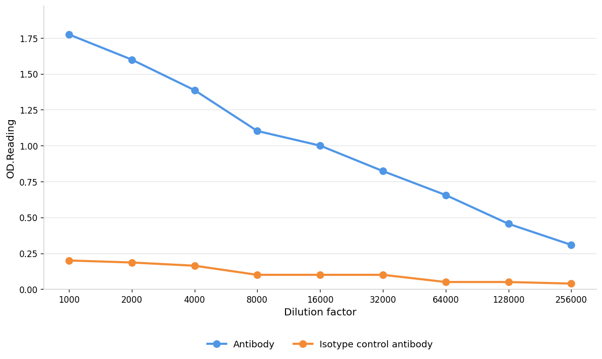
Blue line: Antibody; Orange line: Isotype control antibody| Product Name | Anti-CDK5 antibody,pbs only |
|---|---|
| Antibody Type | Primary Antibodies |
| Immunogen | Recombinant protein |
| Clonality | Monoclonal |
|---|---|
| Isotype | IgG |
| Host Species | Mouse |
| Tested Applications | ELISAICC/IFIF-PIHCWB |
| WB:1:1000 IHC:1:1000 ICC/IF:1:100-1:500 IF-P:1:200-1:2000 |
|
| Species Reactivity | HumanMouseRat |
| Concentration | 1mg/ml |
| Purification | Affinity purified |
| Gene Symbol | CDK5 |
|---|---|
| Gene Synonyms | Crk6 LIS7 PSSALRE |
| Gene Full Name | cyclin dependent kinase 5 |
| Gene Summary | This gene encodes a proline-directed serine/threonine kinase that is a member of the cyclin-dependent kinase family of proteins. Unlike other members of the family, the protein encoded by this gene does not directly control cell cycle regulation. Instead the protein, which is predominantly expressed at high levels in mammalian postmitotic central nervous system neurons, functions in diverse processes such as synaptic plasticity and neuronal migration through phosphorylation of proteins required for cytoskeletal organization, endocytosis and exocytosis, and apoptosis. In humans, an allelic variant of the gene that results in undetectable levels of the protein has been associated with lethal autosomal recessive lissencephaly-7. Alternative splicing results in multiple transcript variants. [provided by RefSeq, May 2015] |
| Alternative Names | CDK5 CDK5 activator 1 CDK5-SV cell division protein kinase 5 CR6 protein kinase cyclin-dependent-like kinase 5 epididymis secretory sperm binding protein protein kinase CDK5 splicing serine/threonine-protein kinase PSSALRE tau protein kinase II catalytic subunit TPKII catalytic subunit |
| Molecular Weight(MW) | 33kDa |
| Cellular Localization | Nucleus cytoplasm |
| SwissProt ID | Q00535 |
|---|
WB
Western blot analysis using CDK5 mouse monoclonal antibody against Hela(1),Hepg2(2),A549(3),Huvec(4),Cos-7(5),P19(6) cell lysate . 12% SDS-PAGE gel.Sample loading: 20μg /lane. Transfer the proteins onto a PVDF membrane (OM790003), and block it with TBST (OM750016) plus skimmed milk powder(OM750084) for one hour. Dilute the primary antibody with the antibody diluent (OM750012) at a ratio of 1:1000, and incubate it overnight at 4°C. Wash the membrane three times with TBST (OM750016), 5 minutes each time. At room temperature, dilute the secondary antibody, Goat Anti-Mouse IgG (H&L) - HRP(OM644366), at a ratio of 1:20000 and incubate for one hour. Wash the membrane three times with TBST (OM750016) again, 5 minutes each time. Use ECL (OM625701) for luminescence.staining time: 60S
IHC
Immunohistochemical analysis of paraffin-embedded breast cancer tissues using CDK5 antibody with DAB staining.Pre-treat the sections with heat-mediated antigen retrieval using sodium citrate buffer (pH 6.0) (OM750020) for 2 minutes. Wash the sections with ddH₂O and PBS (OM750003). Block the tissue with 10% non-immune goat serum(OM760028) at room temperature for 30 minutes. Incubate the tissue with the primary antibody diluted at a ratio of 1:1000 at 4°C overnight. At room temperature, dilute the secondary antibody, Goat Anti-Mouse IgG(H&L)-HRP (OM644366), at a ratio of 1:200 and incubate for one hour. Use DAB(OM760029)as the chromogenic agent. Counterstain the tissue with hematoxylin, and mount the tissue sections with neutral gum.
IHC
Immunohistochemical analysis of paraffin-embedded cervical cancer tissues using CDK5 antibody with DAB staining.Pre-treat the sections with heat-mediated antigen retrieval using sodium citrate buffer (pH 6.0) (OM750020) for 2 minutes. Wash the sections with ddH₂O and PBS (OM750003). Block the tissue with 10% non-immune goat serum(OM760028) at room temperature for 30 minutes. Incubate the tissue with the primary antibody diluted at a ratio of 1:1000 at 4°C overnight. At room temperature, dilute the secondary antibody, Goat Anti-Mouse IgG(H&L)-HRP (OM644366), at a ratio of 1:200 and incubate for one hour. Use DAB(OM760029)as the chromogenic agent. Counterstain the tissue with hematoxylin, and mount the tissue sections with neutral gum.
IHC
Immunohistochemical analysis of paraffin-embedded colon cancer tissues using CDK5 antibody with DAB staining.Pre-treat the sections with heat-mediated antigen retrieval using sodium citrate buffer (pH 6.0) (OM750020) for 2 minutes. Wash the sections with ddH₂O and PBS (OM750003). Block the tissue with 10% non-immune goat serum(OM760028) at room temperature for 30 minutes. Incubate the tissue with the primary antibody diluted at a ratio of 1:1000 at 4°C overnight. At room temperature, dilute the secondary antibody, Goat Anti-Mouse IgG(H&L)-HRP (OM644366), at a ratio of 1:200 and incubate for one hour. Use DAB(OM760029)as the chromogenic agent. Counterstain the tissue with hematoxylin, and mount the tissue sections with neutral gum.
IHC
Immunohistochemical analysis of paraffin-embedded prostate cancer tissues using CDK5 antibody with DAB staining.Pre-treat the sections with heat-mediated antigen retrieval using sodium citrate buffer (pH 6.0) (OM750020) for 2 minutes. Wash the sections with ddH₂O and PBS (OM750003). Block the tissue with 10% non-immune goat serum(OM760028) at room temperature for 30 minutes. Incubate the tissue with the primary antibody diluted at a ratio of 1:1000 at 4°C overnight. At room temperature, dilute the secondary antibody, Goat Anti-Mouse IgG(H&L)-HRP (OM644366), at a ratio of 1:200 and incubate for one hour. Use DAB(OM760029)as the chromogenic agent. Counterstain the tissue with hematoxylin, and mount the tissue sections with neutral gum.
ICC/IF
Immunofluorescence analysis of Hela cells using CDK5 antibody (green). Blue: DAPI fluorescent DNA dye.Cells are fixed in 4% paraformaldehyde at room temperature for 15 minutes. Then, they are permeabilized with a PBS (OM750003) solution containing 0.1% Triton X-100(OM750021) at room temperature for 15 minutes. Subsequently, the cells are blocked with 10% non - immune goat serum(OM760028) at room temperature for 1 hour.The cells are incubated overnight at 4°C with the primary antibody diluted 1:100 in PBS. The secondary antibody, Omnimabs® 488 Goat Ant-Mouse IgG(H&L) (Green,OM643485), is diluted at a ratio of 1:400 and incubated with the cells for 1 hour.Nuclear DNA is labeled with DAPI (Blue,OM643160).
IF-P
Immunohistochemical analysis of paraffin-embedded human cervical cancer tissues using CDK5 antibody with DAB staining.Pre-treat the sections with heat-mediated antigen retrieval using sodium citrate buffer (pH 6.0) (OM750020) for 2 minutes. Wash the sections with ddH₂O and PBS (OM750003). Block the tissue with 10% non-immune goat serum(OM760028) at room temperature for 30 minutes. Incubate the tissue with the primary antibody diluted at a ratio of 1:200 at 4°C overnight. At room temperature, dilute the secondary antibody, Goat Anti-Mouse IgG(H&L)-HRP (OM644366), at a ratio of 1:200 and incubate for one hour. Tyramine labeled with 488 (Green,OM642679) was used as chromogenic agent, and DAPI(Blue,OM642679) was used for double dyeing, and the anti-fluorescence attenuation tablets were sealed.
ELISA
Blue line: Antibody; Orange line: Isotype control antibody| Application Notes | WB:1:1000 IHC:1:1000 ICC/IF:1:100-1:500 IF-P:1:200-1:2000 |
|---|
| Form | Liquid |
|---|---|
| Storage Instructions | Shipped at 4°C. Store at +4°C short term (1-2 weeks). Store at -20°C long term. Avoid freeze / thaw cycle. |
| Storage Buffer | PBS ONLY |


