WB
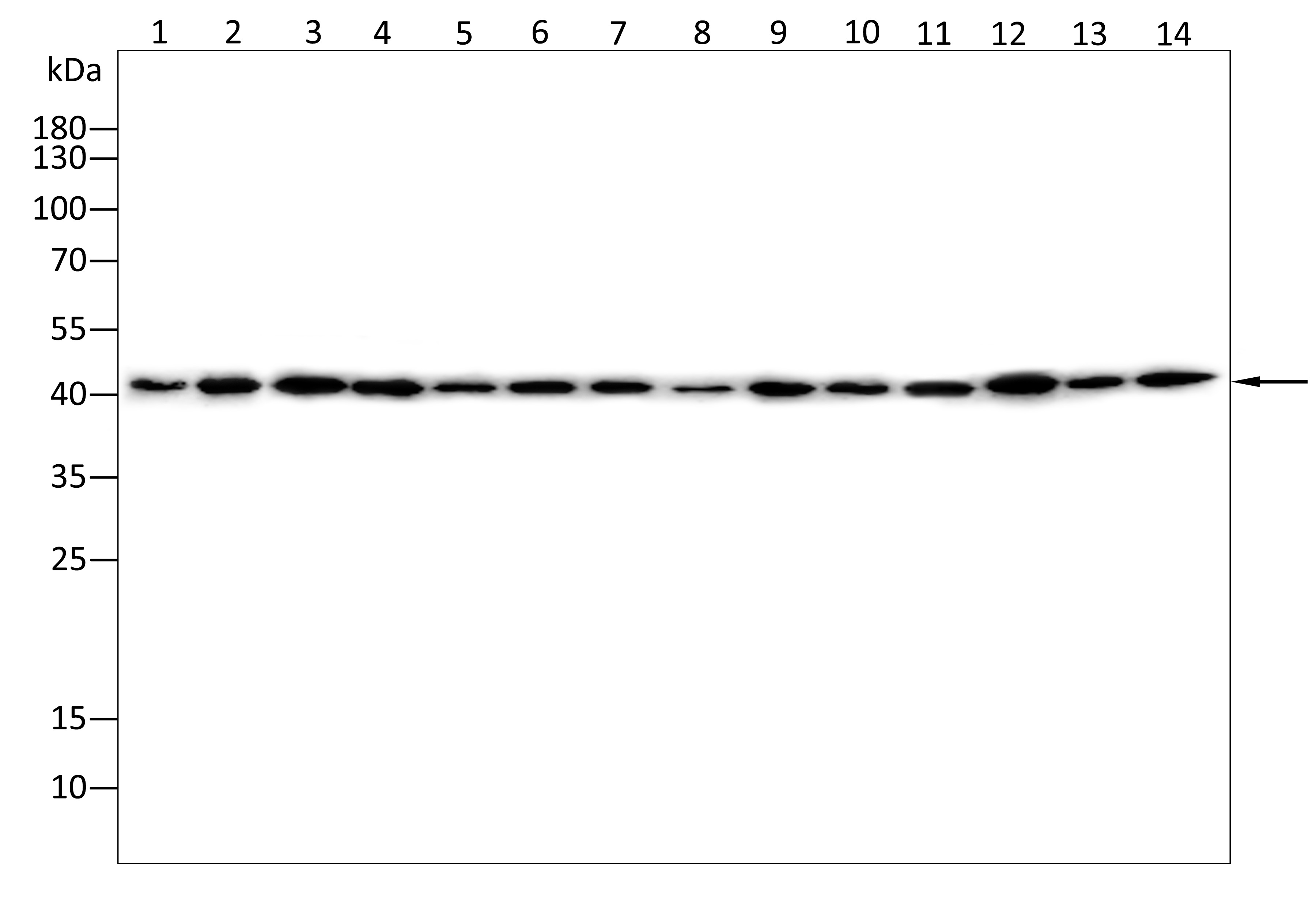
Western blot analysis using ACAT1 antibody against Hela(1),Hepg2(2),K562(3),A549(4),A431(5),Huvec(6),Nih3t3(7),Cos-7(8),C2C12(9),Hek293(10),Mcf-7(11),Pc-12(12),Jurkat(13),Raji(14) cell . 12% SDS-PAGE gel.Sample loading: 20μg /lane. Transfer the proteins onto a PVDF membrane (OM790003), and block it with TBST (OM750016) plus skimmed milk powder(OM750084) for one hour. Dilute the primary antibody with the antibody diluent (OM750012) at a ratio of 1:2000, and incubate it overnight at 4°C. Wash the membrane three times with TBST (OM750016), 5 minutes each time. At room temperature, dilute the secondary antibody, Goat Anti-Rabbit IgG(H&L)-HRP (OM643487), at a ratio of 1:20000 and incubate for one hour. Wash the membrane three times with TBST (OM750016) again, 5 minutes each time. Use ECL (OM625701) for luminescence.staining time: 60S.WB
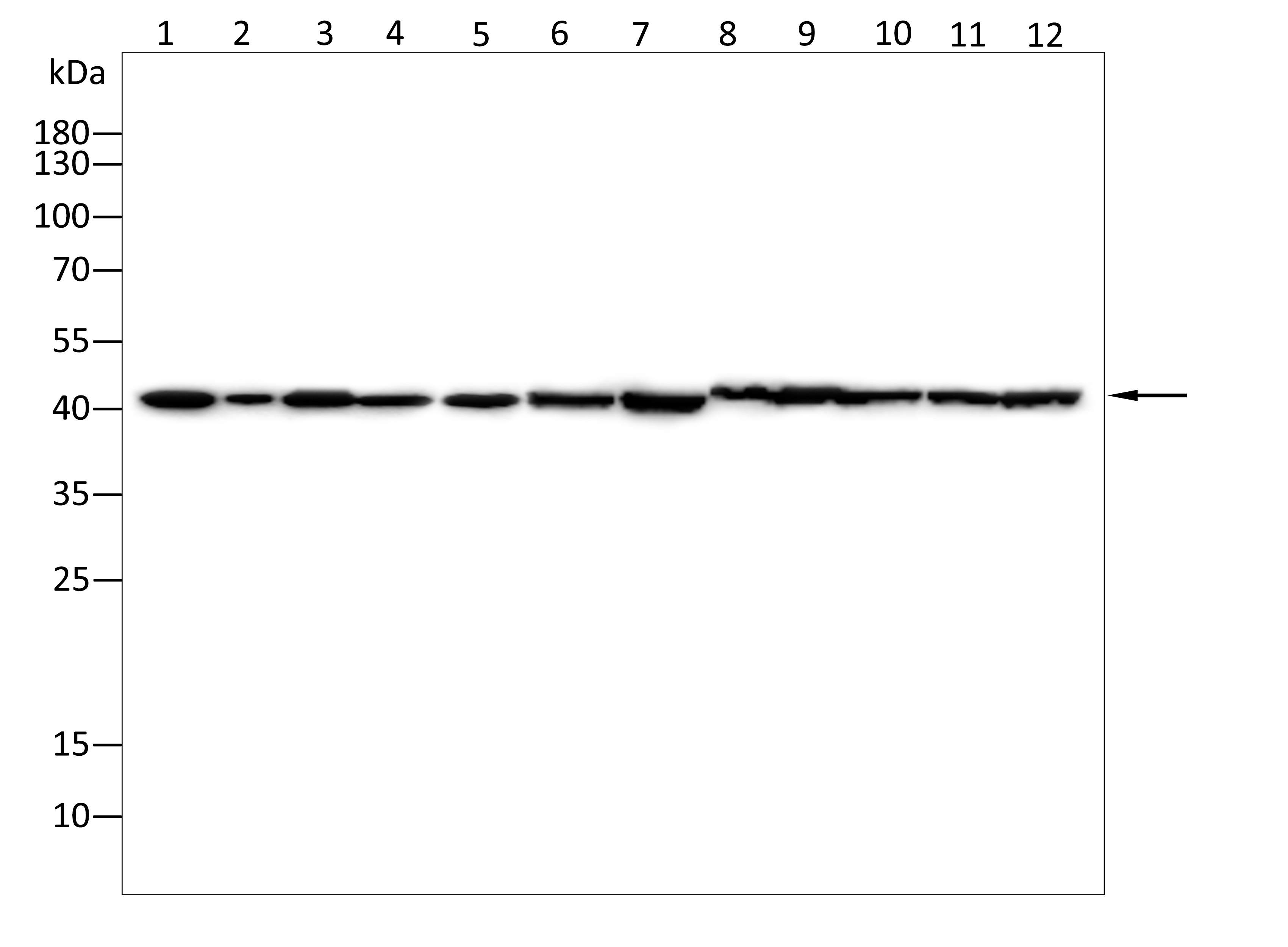
Western blot analysis using ACAT1 antibody against Mouse kidney tissue(1),Mouse brain tissue(2),Mouse liver tissue(3),Mouse lung tissue(4),Mouse spleen tissue(5),Mouse stomach tissue(6),Mouse heart tissue(7),Rat brain tissue(8),Rat liver tissue(9),Rat lung tissue(10), Rat stomach tissue(11),Rat heart tissue(12) . 12% SDS-PAGE gel.Sample loading: 20μg /lane. Transfer the proteins onto a PVDF membrane (OM790003), and block it with TBST (OM750016) plus skimmed milk powder(OM750084) for one hour. Dilute the primary antibody with the antibody diluent (OM750012) at a ratio of 1:2000, and incubate it overnight at 4°C. Wash the membrane three times with TBST (OM750016), 5 minutes each time. At room temperature, dilute the secondary antibody, Goat Anti-Rabbit IgG(H&L)-HRP (OM643487), at a ratio of 1:20000 and incubate for one hour. Wash the membrane three times with TBST (OM750016) again, 5 minutes each time. Use ECL (OM625701) for luminescence.staining time: 60S.ELISA
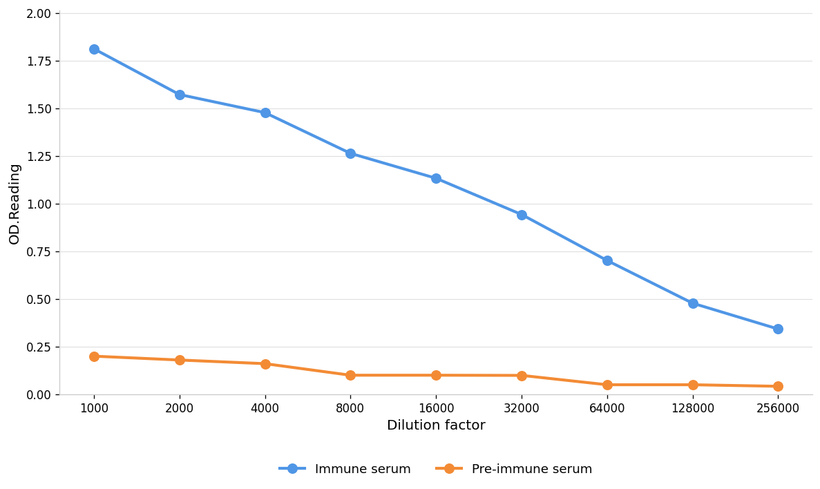
Coat the plate with immunizing antigen.Blue line: Immune serum; Orange line: Pre-immune serum.| Product Name | Anti-ACAT1 antibody,pbs only |
|---|---|
| Antibody Type | Primary Antibodies |
| Immunogen | Polypeptide |
| Clonality | Polyclonal |
|---|---|
| Isotype | IgG |
| Host Species | Rabbit |
| Tested Applications | ELISAWB |
| WB:1:1000-1:2000 |
|
| Species Reactivity | HumanMouseRat |
| Concentration | 1mg/ml |
| Purification | Protein A |
| Gene Symbol | ACAT1 |
|---|---|
| Gene Synonyms | 6330585C21Rik ACAT fd16h07 fd20g06 MAT T2 THIL wu:fd16h07 wu:fd20g06 zgc:86832 |
| Gene Full Name | acetyl-CoA acetyltransferase 1 |
| Gene Summary | This gene encodes a mitochondrially localized enzyme that catalyzes the reversible formation of acetoacetyl-CoA from two molecules of acetyl-CoA. Defects in this gene are associated with 3-ketothiolase deficiency, an inborn error of isoleucine catabolism characterized by urinary excretion of 2-methyl-3-hydroxybutyric acid, 2-methylacetoacetic acid, tiglylglycine, and butanone. [provided by RefSeq, Feb 2009] |
| Alternative Names | acetoacetyl Coenzyme A thiolase acetoacetyl-CoA thiolase acetyl-Coenzyme A acetyltransferase 1 acetyl-Coenzyme A acetyltransferase 1 (acetoacetyl Coenzyme A thiolase) mitochondrial acetoacetyl-CoA thiolase testicular tissue protein Li 198 |
| Molecular Weight(MW) | 45kDa(observed:41kDa) |
| Cellular Localization | Mitochondrion |
| SwissProt ID | P24752 |
|---|
WB
Western blot analysis using ACAT1 antibody against Hela(1),Hepg2(2),K562(3),A549(4),A431(5),Huvec(6),Nih3t3(7),Cos-7(8),C2C12(9),Hek293(10),Mcf-7(11),Pc-12(12),Jurkat(13),Raji(14) cell . 12% SDS-PAGE gel.Sample loading: 20μg /lane. Transfer the proteins onto a PVDF membrane (OM790003), and block it with TBST (OM750016) plus skimmed milk powder(OM750084) for one hour. Dilute the primary antibody with the antibody diluent (OM750012) at a ratio of 1:2000, and incubate it overnight at 4°C. Wash the membrane three times with TBST (OM750016), 5 minutes each time. At room temperature, dilute the secondary antibody, Goat Anti-Rabbit IgG(H&L)-HRP (OM643487), at a ratio of 1:20000 and incubate for one hour. Wash the membrane three times with TBST (OM750016) again, 5 minutes each time. Use ECL (OM625701) for luminescence.staining time: 60S.
WB
Western blot analysis using ACAT1 antibody against Mouse kidney tissue(1),Mouse brain tissue(2),Mouse liver tissue(3),Mouse lung tissue(4),Mouse spleen tissue(5),Mouse stomach tissue(6),Mouse heart tissue(7),Rat brain tissue(8),Rat liver tissue(9),Rat lung tissue(10), Rat stomach tissue(11),Rat heart tissue(12) . 12% SDS-PAGE gel.Sample loading: 20μg /lane. Transfer the proteins onto a PVDF membrane (OM790003), and block it with TBST (OM750016) plus skimmed milk powder(OM750084) for one hour. Dilute the primary antibody with the antibody diluent (OM750012) at a ratio of 1:2000, and incubate it overnight at 4°C. Wash the membrane three times with TBST (OM750016), 5 minutes each time. At room temperature, dilute the secondary antibody, Goat Anti-Rabbit IgG(H&L)-HRP (OM643487), at a ratio of 1:20000 and incubate for one hour. Wash the membrane three times with TBST (OM750016) again, 5 minutes each time. Use ECL (OM625701) for luminescence.staining time: 60S.
ELISA
Coat the plate with immunizing antigen.Blue line: Immune serum; Orange line: Pre-immune serum.| Application Notes | WB:1:1000-1:2000 |
|---|
| Form | Liquid |
|---|---|
| Storage Instructions | Shipped at 4°C. Store at +4°C short term (1-2 weeks). Store at -20°C long term. Avoid freeze / thaw cycle. |
| Storage Buffer | PBS ONLY |


