WB
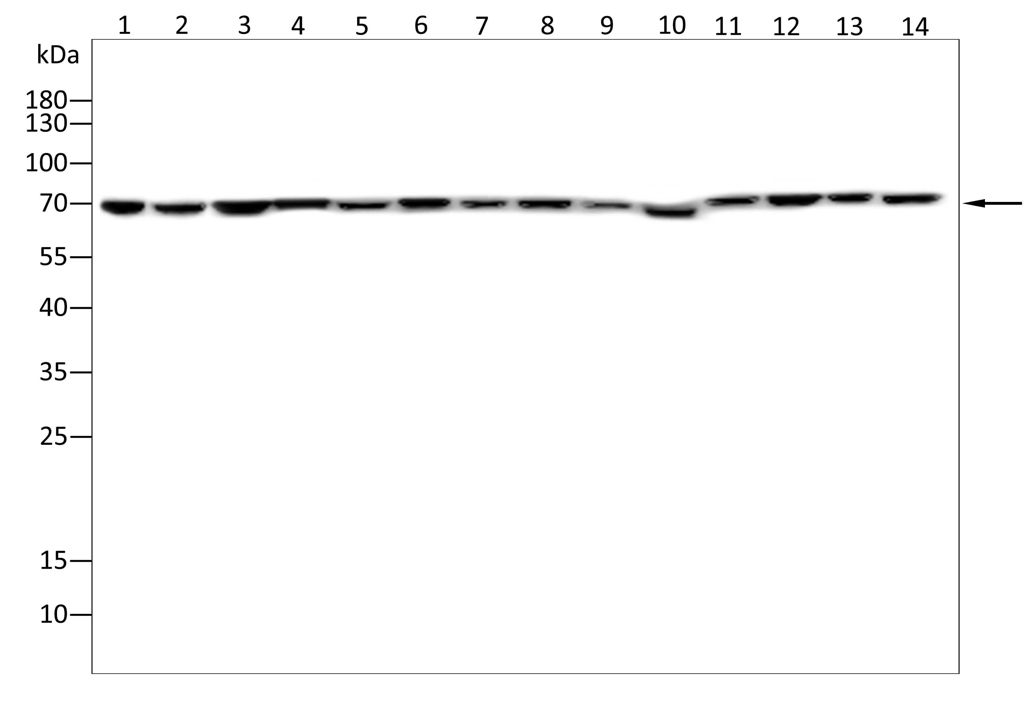
Western blot analysis using LIMPII antibody against Hela(1),Hepg2(2),K562(3),A549(4),A431(5),Huvec(6),Nih3t3(7),Cos-7(8),C2C12(9),Hek293(10),Mcf-7(11),Pc-12(12),Jurkat(13),Raji(14) cell . 12% SDS-PAGE gel.Sample loading: 20μg /lane. Transfer the proteins onto a PVDF membrane (OM790003), and block it with TBST (OM750016) plus skimmed milk powder(OM750084) for one hour. Dilute the primary antibody with the antibody diluent (OM750012) at a ratio of 1:2000, and incubate it overnight at 4°C. Wash the membrane three times with TBST (OM750016), 5 minutes each time. At room temperature, dilute the secondary antibody, Goat Anti-Rabbit IgG(H&L)-HRP (OM643487), at a ratio of 1:20000 and incubate for one hour. Wash the membrane three times with TBST (OM750016) again, 5 minutes each time. Use ECL (OM625701) for luminescence.staining time: 60S.WB
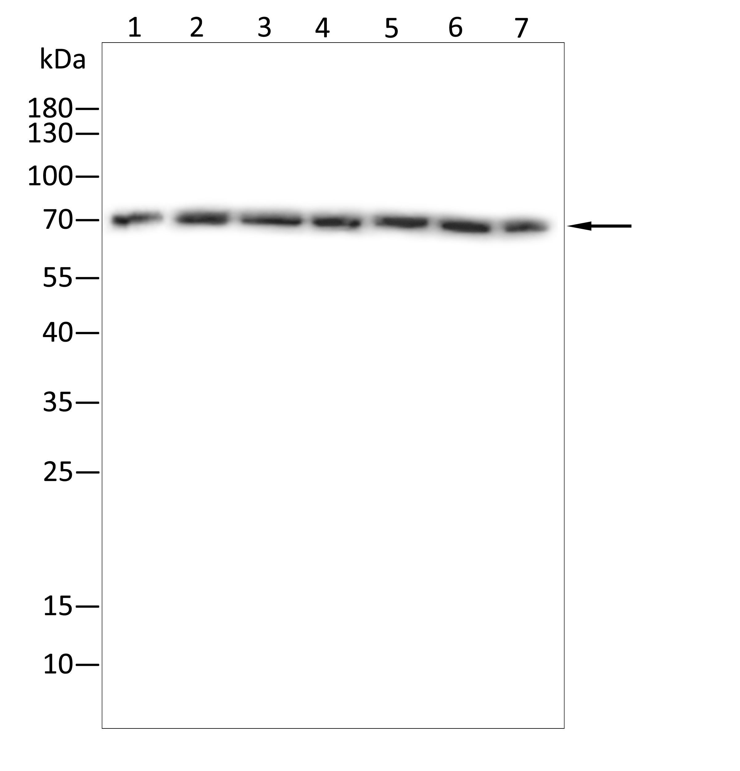
Western blot analysis using LIMPII antibody against Mouse brain tissue(1),Mouse liver tissue(2),Mouse stomach tissue(3),Mouse heart tissue(4),Rat brain tissue(5),Rat liver tissue(6),Rat lung tissue(7) . 12% SDS-PAGE gel.Sample loading: 20μg /lane. Transfer the proteins onto a PVDF membrane (OM790003), and block it with TBST (OM750016) plus skimmed milk powder(OM750084) for one hour. Dilute the primary antibody with the antibody diluent (OM750012) at a ratio of 1:2000, and incubate it overnight at 4°C. Wash the membrane three times with TBST (OM750016), 5 minutes each time. At room temperature, dilute the secondary antibody, Goat Anti-Rabbit IgG(H&L)-HRP (OM643487), at a ratio of 1:20000 and incubate for one hour. Wash the membrane three times with TBST (OM750016) again, 5 minutes each time. Use ECL (OM625701) for luminescence.staining time: 60S.ICC/IF
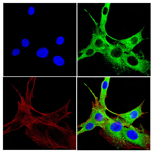
Immunofluorescence analysis of C2C12 cells using LIMPII antibody (green). Blue: DAPI fluorescent DNA dye. Red: Actin filaments have been labeled with Omnimabs® 594-Phalloidin.Cells are fixed in 4% paraformaldehyde at room temperature for 15 minutes. Then, they are permeabilized with a PBS (OM750003) solution containing 0.1% Triton X-100(OM750021) at room temperature for 15 minutes. Subsequently, the cells are blocked with 10% non - immune goat serum(OM760028) at room temperature for 1 hour.The cells are incubated overnight at 4°C with the primary antibody diluted 1:100 in PBS. The secondary antibody, Omnimabs® 488 Goat Ant-Rabbit IgG(H&L) (Green,OM643486), is diluted at a ratio of 1:400 and incubated with the cells for 1 hour.Nuclear DNA is labeled with DAPI (Blue,OM643160). F-actin is stained with Omnimabs® 594-Phalloidin (Red,OM750007) diluted 1:100 for 30 minutes.ELISA
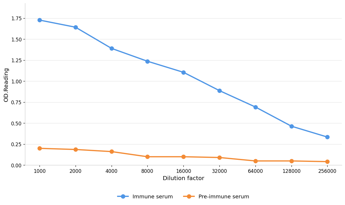
Coat the plate with immunizing antigen.Blue line: Immune serum; Orange line: Pre-immune serum.| Product Name | Anti-LIMPII antibody,pbs only |
|---|---|
| Antibody Type | Primary Antibodies |
| Immunogen | Polypeptide |
| Clonality | Polyclonal |
|---|---|
| Isotype | IgG |
| Host Species | Rabbit |
| Tested Applications | ELISAICC/IFWB |
| WB:1:1000-1:2000 ICC/IF:1:100-1:500 |
|
| Species Reactivity | HumanMouseRat |
| Concentration | 1mg/ml |
| Purification | Protein A |
| Gene Symbol | SCARB2 |
|---|---|
| Gene Synonyms | 9330185J12Rik AMRF CD36L2 EPM4 HLGP85 LGP85 LIMP II LIMP-2 LIMPII MLGP85 SR-BII |
| Gene Full Name | scavenger receptor class B member 2 |
| Gene Summary | The protein encoded by this gene is a type III glycoprotein that is located primarily in limiting membranes of lysosomes and endosomes. Earlier studies in mice and rat suggested that this protein may participate in membrane transportation and the reorganization of endosomal/lysosomal compartment. The protein deficiency in mice was reported to impair cell membrane transport processes and cause pelvic junction obstruction, deafness, and peripheral neuropathy. Further studies in human showed that this protein is a ubiquitously expressed protein and that it is involved in the pathogenesis of HFMD (hand, foot, and mouth disease) caused by enterovirus-71 and possibly by coxsackievirus A16. Mutations in this gene caused an autosomal recessive progressive myoclonic epilepsy-4 (EPM4), also known as action myoclonus-renal failure syndrome (AMRF). Alternatively spliced transcript variants encoding different isoforms have been found for this gene.[provided by RefSeq, Feb 2011] |
| Alternative Names | 85 kDa lysosomal membrane sialoglycoprotein 85 kDa lysosomal sialoglycoprotein scavenger receptor class B member 2 CD 36 CD36 antigen (collagen type I receptor thrombospondin receptor)-like 2 CD36 antigen (collagen type I receptor thrombospondin receptor)-like 2 (lysosomal integral membrane protein II) CD36 antigen-like 2 CD36 molecule LIMP II lysosome membrane protein II sCD 36 sCD36 soluble CD 36 soluble CD36 SR-B2 |
| Molecular Weight(MW) | 54kDa(observed:70kDa) |
| Cellular Localization | Lysosome membrane |
| SwissProt ID | Q14108 |
|---|
WB
Western blot analysis using LIMPII antibody against Hela(1),Hepg2(2),K562(3),A549(4),A431(5),Huvec(6),Nih3t3(7),Cos-7(8),C2C12(9),Hek293(10),Mcf-7(11),Pc-12(12),Jurkat(13),Raji(14) cell . 12% SDS-PAGE gel.Sample loading: 20μg /lane. Transfer the proteins onto a PVDF membrane (OM790003), and block it with TBST (OM750016) plus skimmed milk powder(OM750084) for one hour. Dilute the primary antibody with the antibody diluent (OM750012) at a ratio of 1:2000, and incubate it overnight at 4°C. Wash the membrane three times with TBST (OM750016), 5 minutes each time. At room temperature, dilute the secondary antibody, Goat Anti-Rabbit IgG(H&L)-HRP (OM643487), at a ratio of 1:20000 and incubate for one hour. Wash the membrane three times with TBST (OM750016) again, 5 minutes each time. Use ECL (OM625701) for luminescence.staining time: 60S.
WB
Western blot analysis using LIMPII antibody against Mouse brain tissue(1),Mouse liver tissue(2),Mouse stomach tissue(3),Mouse heart tissue(4),Rat brain tissue(5),Rat liver tissue(6),Rat lung tissue(7) . 12% SDS-PAGE gel.Sample loading: 20μg /lane. Transfer the proteins onto a PVDF membrane (OM790003), and block it with TBST (OM750016) plus skimmed milk powder(OM750084) for one hour. Dilute the primary antibody with the antibody diluent (OM750012) at a ratio of 1:2000, and incubate it overnight at 4°C. Wash the membrane three times with TBST (OM750016), 5 minutes each time. At room temperature, dilute the secondary antibody, Goat Anti-Rabbit IgG(H&L)-HRP (OM643487), at a ratio of 1:20000 and incubate for one hour. Wash the membrane three times with TBST (OM750016) again, 5 minutes each time. Use ECL (OM625701) for luminescence.staining time: 60S.
ICC/IF
Immunofluorescence analysis of C2C12 cells using LIMPII antibody (green). Blue: DAPI fluorescent DNA dye. Red: Actin filaments have been labeled with Omnimabs® 594-Phalloidin.Cells are fixed in 4% paraformaldehyde at room temperature for 15 minutes. Then, they are permeabilized with a PBS (OM750003) solution containing 0.1% Triton X-100(OM750021) at room temperature for 15 minutes. Subsequently, the cells are blocked with 10% non - immune goat serum(OM760028) at room temperature for 1 hour.The cells are incubated overnight at 4°C with the primary antibody diluted 1:100 in PBS. The secondary antibody, Omnimabs® 488 Goat Ant-Rabbit IgG(H&L) (Green,OM643486), is diluted at a ratio of 1:400 and incubated with the cells for 1 hour.Nuclear DNA is labeled with DAPI (Blue,OM643160). F-actin is stained with Omnimabs® 594-Phalloidin (Red,OM750007) diluted 1:100 for 30 minutes.
ELISA
Coat the plate with immunizing antigen.Blue line: Immune serum; Orange line: Pre-immune serum.| Application Notes | WB:1:1000-1:2000 ICC/IF:1:100-1:500 |
|---|
| Form | Liquid |
|---|---|
| Storage Instructions | Shipped at 4°C. Store at +4°C short term (1-2 weeks). Store at -20°C long term. Avoid freeze / thaw cycle. |
| Storage Buffer | PBS ONLY |


