Immunohistochemical
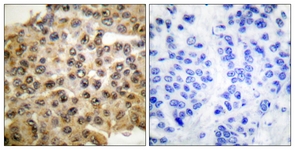
Immunohistochemistry analysis of paraffin-embedded human breast carcinoma tissue, using p44/42 MAPK Antibody. The picture on the right is treated with the synthesized peptide.Western blot
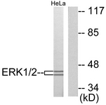
Western blot analysis of extracts from HeLa cells, using p44/42 MAPK Antibody. The lane on the right is treated with the synthesized peptideWB
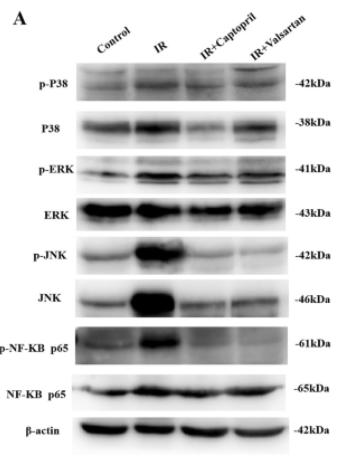
(A) Expression of MAPKs and NF-κB in the lung tissues.WB
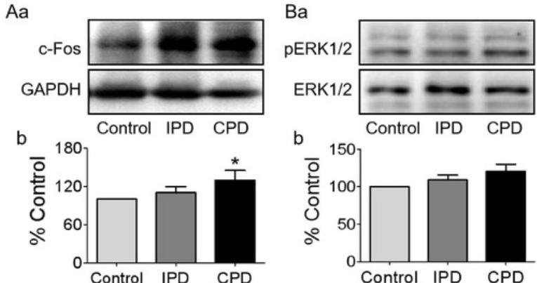
(A and B) Western blotting bands (a) and their statistical analyses (b) of c-Fos (A, n = 6) and pERK1/2 (B, n = 7).WB
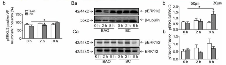
(B and C) Western blotting bands (a) and the summary graphs (b) showing the effects of BAO and BC on pERK1/2 expression in SON (B) and PVN (C).WB
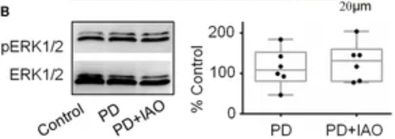
(B) Western blot bands (left panel) and the summary graphs (right panel) showing the expression of pERK1/2 under different experimental conditions.WB
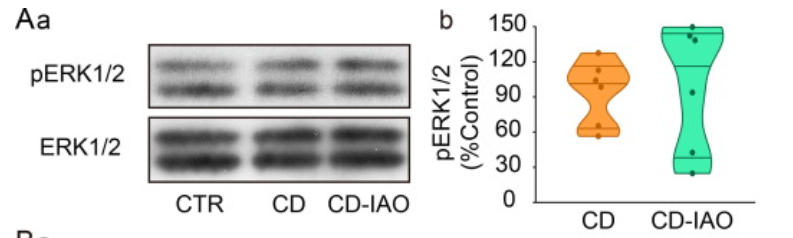
(A) Representative Western blotting bands (a) and the summary graph (b) showing the effects of CD and of CD-IAO on phosphorylated extracellular signal-regulated protein kinase 1/2 (pERK 1/2) protein expression.WB
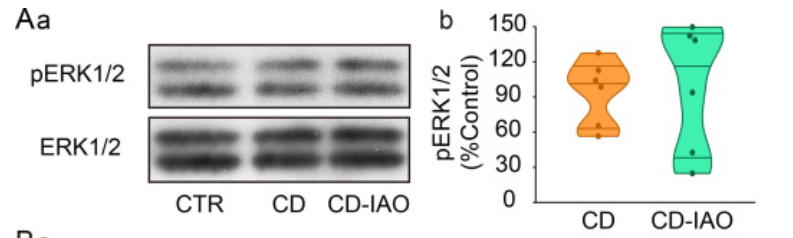
(A) Representative Western blotting bands (a) and the summary graph (b) showing the effects of CD and of CD-IAO on pERK 1/2 protein expression.| Product Name | Rabbit anti-p44/42 MAPK polyclonal antibody |
|---|---|
| Antibody Type | Primary Antibodies |
| Immunogen | The antiserum was produced against synthesized peptide derived from human p44/42 MAPK. |
| Isotype | Cocktail |
|---|---|
| Host Species | Rabbit |
| Tested Applications | ELISAICC/IFIHCWB |
| WB:1:500~1:1000 IHC:1:50~1:100 ELISA:1:10000 1:200: |
|
| Species Reactivity | HumanMouseRat |
| Concentration | 1mg/ml |
| Purification | Affinity purified |
| Gene Symbol | MAPK3 |
|---|---|
| Gene Synonyms | ERK-1 ERK1 ERT2 Extracellular signal- regulated kinase 1 Insulin-stimulated MAP2 kinase kinase ERK1 MAP kinase 1 MAPK 1 MAPK3 Microtubule- associated protein-2 kinase Mitogen-activated protein kinase 3 MK03 MNK1 p44-ERK1 P44-ERK1 p44-MAPK P |
Immunohistochemical
Immunohistochemistry analysis of paraffin-embedded human breast carcinoma tissue, using p44/42 MAPK Antibody. The picture on the right is treated with the synthesized peptide.
Western blot
Western blot analysis of extracts from HeLa cells, using p44/42 MAPK Antibody. The lane on the right is treated with the synthesized peptide
WB
(A) Expression of MAPKs and NF-κB in the lung tissues.
WB
(A and B) Western blotting bands (a) and their statistical analyses (b) of c-Fos (A, n = 6) and pERK1/2 (B, n = 7).
WB
(B and C) Western blotting bands (a) and the summary graphs (b) showing the effects of BAO and BC on pERK1/2 expression in SON (B) and PVN (C).
WB
(B) Western blot bands (left panel) and the summary graphs (right panel) showing the expression of pERK1/2 under different experimental conditions.
WB
(A) Representative Western blotting bands (a) and the summary graph (b) showing the effects of CD and of CD-IAO on phosphorylated extracellular signal-regulated protein kinase 1/2 (pERK 1/2) protein expression.
WB
(A) Representative Western blotting bands (a) and the summary graph (b) showing the effects of CD and of CD-IAO on pERK 1/2 protein expression.| Application Notes | WB:1:500~1:1000 IHC:1:50~1:100 ELISA:1:10000 1:200: |
|---|
| Form | Liquid |
|---|---|
| Storage Instructions | store at -20°C. Avoid repeated freeze / thaw cycles |
| Storage Buffer | Rabbit IgG in phosphate buffered saline (without Mg2+ and Ca2+), pH 7.4, 150mM NaCl, 0.02% sodium azide and 50% glycerol. |


