Application
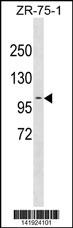
PDCD6IP Antibody (Center) western blot analysis in ZR-75-1 cell line lysates (35ug/lane).This demonstrates the PDCD6IP antibody detected the PDCD6IP protein (arrow).| Product Name | PDCD6IP Antibody (Center) |
|---|---|
| Antibody Type | Primary Antibodies |
| Antigen Alias | PDCD6IP; AIP1; ALIX; KIAA1375; Programmed cell death 6-interacting protein; ALG-2-interacting protein 1; Hp95 |
| Clonality | Polyclonal |
|---|---|
| Isotype | Ig |
| Host Species | Rabbit |
| Tested Applications | WB |
| WB:1:100~500 |
|
| Species Reactivity | Human |
| Concentration | 1mg/ml |
| Gene Synonyms | AIP1 ALIX KIAA1375 |
|---|---|
| Alternative Names | PDCD6IP AIP1 ALIX KIAA1375 Programmed cell death 6-interacting protein ALG-2-interacting protein 1 Hp95 |
| Molecular Weight(MW) | 96023 Da |
| Function | Class E VPS protein involved in concentration and sorting of cargo proteins of the multivesicular body (MVB) for incorporation into intralumenal vesicles (ILVs) that are generated by invagination and scission from the limiting membrane of the endosome. Binds to the phospholipid lysobisphosphatidic acid (LBPA) which is abundant in MVBs internal membranes. The MVB pathway appears to require the sequential function of ESCRT-O, -I,-II and -III complexes. The ESCRT machinery also functions in topologically equivalent membrane fission events, such as the terminal stages of cytokinesis and enveloped virus budding (HIV-1 and other lentiviruses). Appears to be an adapter for a subset of ESCRT-III proteins, such as CHMP4, to function at distinct membranes. Required for completion of cytokinesis. Involved in HIV-1 virus budding. Can replace TSG101 it its role of supporting HIV-1 release; this function implies the interaction with CHMP4B May play a role in the regulation of both apoptosis and cell proliferation |
| Tissue Specificity | This PDCD6IP antibody is generated from rabbits immunized with a KLH conjugated synthetic peptide between 541-570 amino acids from the Central region of human PDCD6IP. |
| Cellular Localization | Cytoplasm, cytosol. Melanosome. Cytoplasm, cytoskeleton, centrosome. Note=Identified by mass spectrometry in melanosome fractions from stage I to stage IV. Colocalized with CEP55 in the midbody during cytokinesis. Colocalized with CEP55 at centrosomes of non-dividing cells |
| Entrez Gene | 10015 |
|---|---|
| Nucleotide Accession | NP_037506.2 |
Application
PDCD6IP Antibody (Center) western blot analysis in ZR-75-1 cell line lysates (35ug/lane).This demonstrates the PDCD6IP antibody detected the PDCD6IP protein (arrow).| Application Notes | WB:1:100~500 |
|---|
| Form | Liquid |
|---|---|
| Storage Instructions | For short-term storage, store at 4° C. For long-term storage, aliquot and store at -20ºC or below. Avoid multiple freeze-thaw cycles. |
| Storage Buffer | Purified polyclonal antibody supplied in PBS with 0.09% (W/V) sodium azide. This antibody is purified through a protein A column, followed by peptide affinity purification. |


