Application
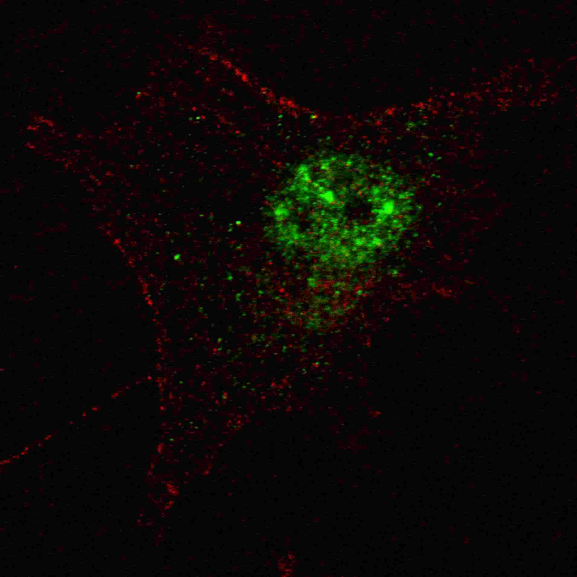
Fluorescent confocal image of SY5Y cells stained with PDX1 (T11) antibody. SY5Y cells were fixed with 4% PFA (20 min), permeabilized with Triton X-100 (0.2%, 30 min). Cells were then incubated with AP2034d PDX1 (T11) primary antibody (1:100, 2 h at room temperature). For secondary antibody, Alexa Fluor® 488 conjugated donkey anti-rabbit antibody (green) was used (1:1000, 1h). Nuclei were counterstained with Hoechst 33342 (blue) (10 μg/ml, 5 min). Note the highly specific localization of the PDX1 immunosignal to the nucleus, supported by Human Protein Atlas Data (http://www.proteinatlas.org/ENSG00000110435).Application
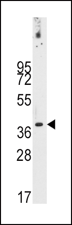
Western blot analysis of anti-PDX1 Antibody (T11) in NCI-H460 cell line lysates (35ug/lane).PDX1-T11(arrow) was detected using the purified Pab.Application
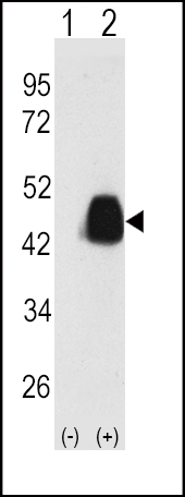
Western blot analysis of PDX1 (arrow) using PDX1 Antibody (T11) . 293 cell lysates (2 ug/lane) either nontransfected (Lane 1) or transiently transfected with the PDX1 gene (Lane 2) (Origene Technologies).Application
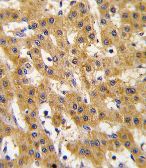
Formalin-fixed and paraffin-embedded human hepatocarcinoma tissue reacted with Phospho-PDX1-T11.ctrl antibody, which was peroxidase-conjugated to the secondary antibody, followed by DAB staining. This data demonstrates the use of this antibody for immunohistochemistry; clinical relevance has not been evaluated.| Product Name | PDX1 Antibody (T11) |
|---|---|
| Antibody Type | Primary Antibodies |
| Antigen Alias | PDHX; PDX1; Pyruvate dehydrogenase protein X component, mitochondrial; Dihydrolipoamide dehydrogenase-binding protein of pyruvate dehydrogenase complex; E3-binding protein; Lipoyl-containing pyruvate dehydrogenase complex component X; proX |
| Clonality | Polyclonal |
|---|---|
| Isotype | Ig |
| Host Species | Rabbit |
| Tested Applications | IFWBIHC |
| IF:1:100 WB |
|
| Species Reactivity | Human |
| Concentration | 1mg/ml |
| Gene Synonyms | PDX1 |
|---|---|
| Alternative Names | PDHX PDX1 Pyruvate dehydrogenase protein X component mitochondrial Dihydrolipoamide dehydrogenase-binding protein of pyruvate dehydrogenase complex E3-binding protein Lipoyl-containing pyruvate dehydrogenase complex component X proX |
| Molecular Weight(MW) | 54122 Da |
| Function | Required for anchoring dihydrolipoamide dehydrogenase (E3) to the dihydrolipoamide transacetylase (E2) core of the pyruvate dehydrogenase complexes of eukaryotes. This specific binding is essential for a functional PDH complex |
| Tissue Specificity | This PDX1 antibody is generated from rabbits immunized with a KLH conjugated synthetic peptide between 1~30 amino acids surrounding T11 of human PDX1. |
| Cellular Localization | Mitochondrion matrix. |
| Entrez Gene | 8050 |
|---|
Application
Fluorescent confocal image of SY5Y cells stained with PDX1 (T11) antibody. SY5Y cells were fixed with 4% PFA (20 min), permeabilized with Triton X-100 (0.2%, 30 min). Cells were then incubated with AP2034d PDX1 (T11) primary antibody (1:100, 2 h at room temperature). For secondary antibody, Alexa Fluor® 488 conjugated donkey anti-rabbit antibody (green) was used (1:1000, 1h). Nuclei were counterstained with Hoechst 33342 (blue) (10 μg/ml, 5 min). Note the highly specific localization of the PDX1 immunosignal to the nucleus, supported by Human Protein Atlas Data (http://www.proteinatlas.org/ENSG00000110435).
Application
Western blot analysis of anti-PDX1 Antibody (T11) in NCI-H460 cell line lysates (35ug/lane).PDX1-T11(arrow) was detected using the purified Pab.
Application
Western blot analysis of PDX1 (arrow) using PDX1 Antibody (T11) . 293 cell lysates (2 ug/lane) either nontransfected (Lane 1) or transiently transfected with the PDX1 gene (Lane 2) (Origene Technologies).
Application
Formalin-fixed and paraffin-embedded human hepatocarcinoma tissue reacted with Phospho-PDX1-T11.ctrl antibody, which was peroxidase-conjugated to the secondary antibody, followed by DAB staining. This data demonstrates the use of this antibody for immunohistochemistry; clinical relevance has not been evaluated.| Application Notes | IF:1:100 WB |
|---|
| Form | Liquid |
|---|---|
| Storage Instructions | For short-term storage, store at 4° C. For long-term storage, aliquot and store at -20ºC or below. Avoid multiple freeze-thaw cycles. |
| Storage Buffer | Purified polyclonal antibody supplied in PBS with 0.09% (W/V) sodium azide. This antibody is purified through a protein A column, followed by peptide affinity purification. |


